Clear Sky Science · en
Construction of a diagnostic model for preeclampsia based on differentially expressed lactylation-related genes and the immune infiltration analysis
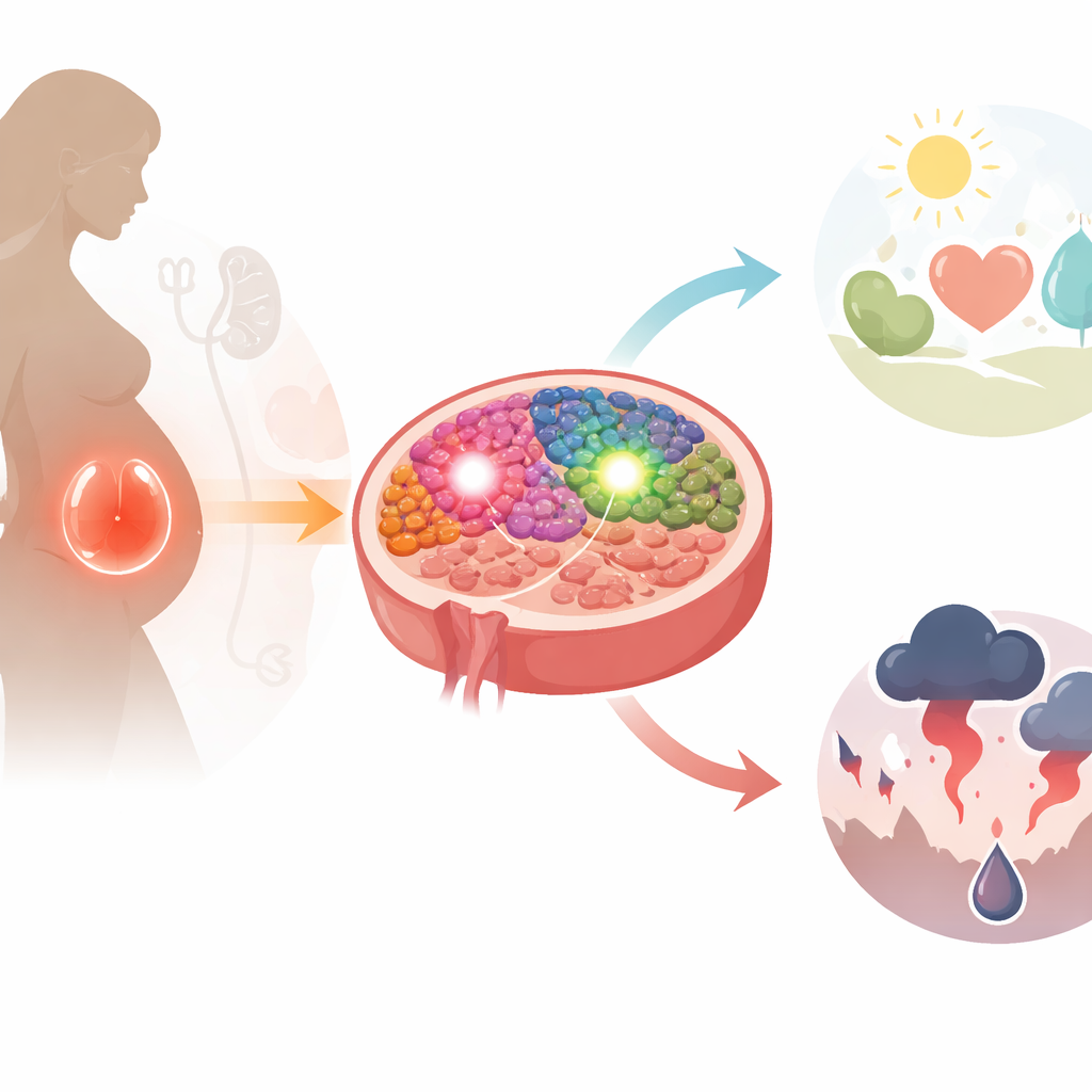
Why this pregnancy research matters
Preeclampsia is a dangerous complication of pregnancy that raises blood pressure and can damage organs in both mother and baby. Doctors today can usually only act once symptoms have appeared, often by delivering the baby early. This study asks a pressing question: can we read early warning signs in the placenta’s genes and cells, long before preeclampsia becomes life‑threatening? By tracing how certain metabolic changes and immune cells behave in the placenta, the authors search for simple genetic signals that might one day help predict and better manage this condition.
Looking for genetic warning lights
The researchers began by gathering several large public datasets of placental tissue from women with and without preeclampsia. They searched these datasets for genes that were switched on or off differently in preeclampsia. From nearly a hundred such genes, they focused on a special subset tied to a recently discovered protein modification called lactylation, in which a small chemical tag derived from lactate is added to proteins. Two genes stood out: EAF1 and PFKP. Both were consistently more active in placentas from women with preeclampsia than in healthy controls, suggesting that they might act as “warning lights” for the disease.
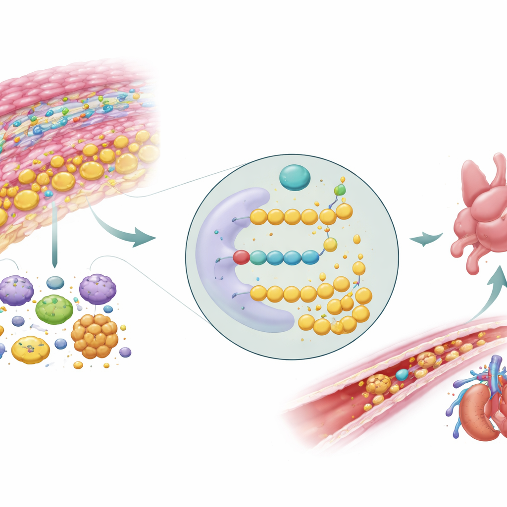
Energy use and a stressed placenta
When the team examined what these two genes do, they found that they were closely linked to how cells process sugar for energy. In particular, the genes were enriched in pathways related to glycolysis, the fast, oxygen‑poor way cells break down glucose, and to the related gluconeogenesis pathway. PFKP is a key enzyme that helps control the speed of glycolysis, while EAF1 influences how other genes are turned on. In preeclampsia, the placenta is often starved of oxygen, which pushes cells to rely more on glycolysis and to generate more lactate. The study’s analyses show that gene activity patterns connected to EAF1 and PFKP differ sharply between preeclampsia and normal pregnancies, reinforcing the idea that disrupted energy use is central to this disorder.
Building a predictive model from two genes
Using EAF1 and PFKP together, the authors built a statistical model to distinguish placentas from women with preeclampsia and those from healthy pregnancies. In the main group of over 200 samples, this two‑gene model correctly classified about four out of five cases. When they tested it on an entirely separate dataset, its accuracy rose to more than nine out of ten samples. Additional checks showed that the model’s predictions closely matched real outcomes and that, in simulated clinical scenarios, using the model would provide more benefit than simple “treat all” or “treat none” strategies. The team then confirmed, using placental tissue collected at their own hospital, that both EAF1 and PFKP were indeed more abundant at the RNA level in preeclampsia samples.
Immune cells and hidden subtypes
Preeclampsia is not just a problem of blood vessels and hormones; it also alters the immune landscape of the placenta. By applying a computational tool that estimates the mix of immune cells in tissue, the researchers found that plasma cells, killer T cells, regulatory T cells, activated dendritic cells, and activated mast cells were more common in preeclampsia, whereas certain helper T cells, natural killer cells, monocytes, and anti‑inflammatory macrophages were relatively depleted. Using patterns of EAF1 and PFKP activity, they further divided preeclampsia cases into two molecular subtypes that differed in their immune cell profiles, especially in the levels of activated dendritic cells. This hints that what clinicians call “preeclampsia” may in fact encompass biologically distinct forms that could respond differently to treatment.
Zooming in on single cells in the placenta
To understand where these genes are active, the team turned to single‑cell RNA sequencing, a technique that reads gene activity in thousands of individual cells. They mapped more than a dozen cell types in the placenta, including different trophoblast cells that form the interface between mother and fetus, immune cells such as macrophages and Hofbauer cells, and developing blood cells. EAF1 was most strongly expressed in certain macrophages and extravillous trophoblast cells, while PFKP was concentrated in extravillous trophoblasts. Many of these cell types showed altered proportions in preeclampsia, with some trophoblast and B‑cell populations expanded and several macrophage‑like cells reduced. Together, these shifts suggest that EAF1 and PFKP are woven into a complex network of energy metabolism and immune regulation in specific cells that help anchor and nourish the placenta.
What this means for future pregnancy care
In plain terms, this study proposes that just two genes involved in how placental cells burn sugar and handle lactate may serve as promising markers for preeclampsia. By combining big‑data analysis with fine‑grained single‑cell views, the authors link these genes to disturbed energy use, a skewed immune environment, and distinct subtypes of the disease. While larger and more diverse clinical studies are still needed before any test reaches the clinic, the work points toward a future in which a simple gene‑based panel, informed by how the placenta makes and uses energy, could help doctors spot high‑risk pregnancies earlier and tailor monitoring and treatment more precisely.
Citation: Zhang, J., Peng, Q., Fei, K. et al. Construction of a diagnostic model for preeclampsia based on differentially expressed lactylation-related genes and the immune infiltration analysis. Sci Rep 16, 14471 (2026). https://doi.org/10.1038/s41598-026-45138-4
Keywords: preeclampsia, placenta, lactylation, glycolysis, immune cells