Clear Sky Science · en
Single-nucleus ATAC-seq analysis resolves chromatin and transcriptional features of fibrolamellar carcinoma
Peering Into a Rare Liver Cancer
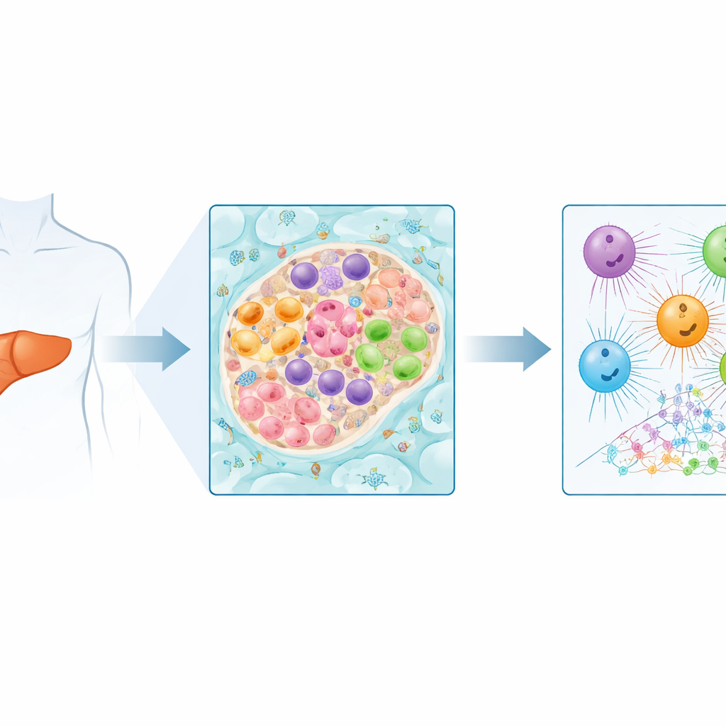
Fibrolamellar carcinoma is a rare form of liver cancer that most often strikes teenagers and young adults, and for many patients surgery is the only hope. Yet this tumor is not made of cancer cells alone: it is a complex community of many cell types interacting in a shared environment. This study uses cutting-edge single-cell technologies to zoom in on that community, revealing which cells drive key molecular changes and pointing toward new ways to target the disease.
Why This Cancer Is So Challenging
Fibrolamellar carcinoma stands apart from more common liver cancers. It typically arises in people without underlying liver disease and is marked by thick bands of scar-like tissue running through the tumor. Almost all patients share a distinctive DNA fusion that creates an abnormal protein, DNAJ-PKAc, which can kick-start tumors in animal models. But directly blocking this protein has proven difficult because similar proteins are essential for normal cell function. As a result, researchers increasingly focus on the downstream pathways and cell types that this fusion protein disrupts, in hopes of finding more precise points of attack.
Looking at Each Cell One by One
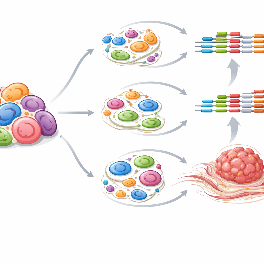
Previous large-scale studies measured gene activity and chromatin activity (how tightly DNA is packaged) by blending all the cells from a tumor together. That approach is powerful but masks the roles of individual cell types such as tumor cells, blood vessel cells, and scar-forming support cells. In this work, the authors isolate individual nuclei from frozen tumor samples and nearby non-cancerous liver tissue. They apply single-nucleus ATAC-seq, which maps open regions of DNA where regulatory elements reside, and single-nucleus RNA-seq, which measures which genes are turned on. Sophisticated computational methods group nuclei into clusters that match known liver cell types and compare how those cells differ between tumor and normal tissue.
Different Cells, Different Molecular Jobs
The single-cell view reveals that many previously reported molecular signals in fibrolamellar carcinoma actually arise from distinct cell types within the tumor. For example, three small regulatory RNAs once thought of simply as tumor markers show sharply different patterns: miR-190b activity concentrates in cancerous liver epithelial cells, miR-10b is mainly altered in blood vessel cells, and miR-199b is elevated in activated stellate cells, which are key players in scarring. This means that simple cancer cell models are not sufficient to study all of these molecules. The team also maps DNA regions where certain transcription factors, the proteins that control gene activity, are likely to bind. Networks involving AP-1 family members and CREB proteins appear rewired in specific cell types, while signals linked to normal liver function are diminished in tumor cells.
Hidden Control Switches and Tumor Scarring
By integrating their single-cell chromatin data with earlier bulk measurements of active regulatory elements, the researchers assign powerful “super enhancers” to particular cell types. Many of these DNA control hubs lie near genes that are unusually active in fibrolamellar carcinoma. One highlighted example is CDH11, a gene previously linked to fibrosis and immune suppression in other diseases. The study finds that distinct super enhancers near CDH11 are active in both tumor cells and activated stellate cells, and that CDH11 itself is expressed in both. Additional analyses of cell-to-cell communication suggest that stellate cells act as hubs for TGFβ signaling, a pathway known to drive scarring. Together, this points to a converging network in which CDH11 and certain microRNAs help fuel the formation of the thick fibrous tissue that characterizes this cancer.
What This Means for Future Treatments
This work delivers a high-resolution map of the regulatory landscape in fibrolamellar carcinoma, broken down by cell type. For non-specialists, the key message is that the tumor’s behavior cannot be explained by cancer cells alone; supporting cells and blood vessels play crucial roles, and different molecules act in different cellular “neighborhoods.” By identifying which genes, control switches, and signaling pathways are active in which cells, the study highlights candidates such as CDH11, specific microRNAs, and transcription factor networks that might be targeted more safely and effectively than the original fusion protein itself. While further validation and more patient-matched samples are needed, this single-cell atlas provides a valuable guide for designing future therapies for this rare and formidable liver cancer.
Citation: Farghli, A.R., Sherman, M.S., Shui, B. et al. Single-nucleus ATAC-seq analysis resolves chromatin and transcriptional features of fibrolamellar carcinoma. Sci Rep 16, 14360 (2026). https://doi.org/10.1038/s41598-026-44899-2
Keywords: fibrolamellar carcinoma, single-cell sequencing, chromatin accessibility, tumor microenvironment, liver cancer