Clear Sky Science · en
PTP4A3 contributes to pathological retinal neovascularization and vascular leakage partly through the PI3K-AKT signalling pathway
Why this matters for people with diabetes
For millions of people living with diabetes, one of the most feared complications is losing vision. Diabetic eye disease damages the fine blood vessels that nourish the light‑sensing tissue at the back of the eye, the retina. This study explores a lesser‑known molecule inside those vessels, called PTP4A3, and shows that it plays a surprising role in making retinal vessels both leakier and more likely to sprout fragile new branches. By targeting this molecule, the researchers suggest, we may one day complement or even improve on current eye injections that do not work well for everyone.
A closer look at diabetic eye damage
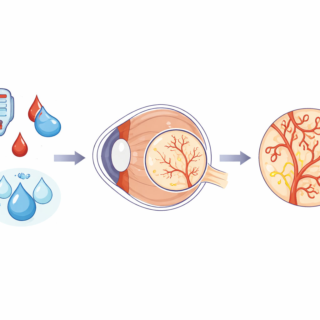
Diabetic retinopathy develops when years of high blood sugar and poor oxygen supply injure the delicate lining of retinal blood vessels. Normally, these vessels form a tight, well‑sealed network that keeps fluid where it belongs. In advanced disease, however, they begin to leak and to grow abnormal new branches, a process called pathological neovascularization. Today’s mainstay treatment involves repeated injections that block a growth signal known as VEGF, yet up to a quarter of patients respond poorly and many continue to have swelling in the central retina. This gap in care has driven scientists to search for additional culprits beyond VEGF that could be targeted to better preserve sight.
A hidden switch that goes into overdrive
The team focused on PTP4A3, an enzyme better known for its role in cancer, where it helps tumor cells grow and spread and encourages new blood vessel formation. Using human eye tissue data and two well‑established mouse models that mimic forms of diabetic eye disease, they found that PTP4A3 levels were markedly higher whenever retinas showed the kind of leaky, overgrown vessels seen in severe diabetic retinopathy. They also recreated diabetes‑like stress in cultured retinal vessel cells by exposing them to high sugar and low oxygen, and again saw PTP4A3 levels climb. Together, these observations pointed to PTP4A3 as an overactive internal “switch” in damaged retinal vessels.
How this switch harms retinal blood vessels
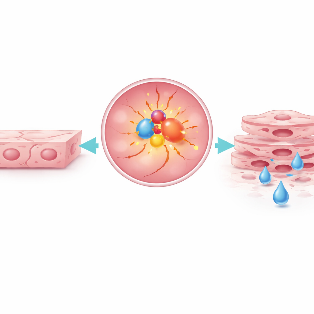
To learn what this switch actually does, the researchers artificially boosted PTP4A3 inside retinal vessel cells. The cells began to divide faster and migrate more readily—exactly the behaviors that drive unwanted vessel sprouting in diseased retinas. Just as importantly, the normally tight barrier between neighboring cells weakened. Key sealing proteins that act like rivets between cells, such as occludin and claudin‑5, were reduced, and lab tests showed more dye slipping through the cell layer. Interestingly, this leakiness did not come from extra transport through the cells themselves but from loosened junctions between them, turning the vessel wall from a watertight hose into a sieve.
Dialing down the damage with targeted blockers
The study then asked whether turning this switch back down could protect the retina. In cells, a small‑molecule drug that blocks PTP4A3 reversed many of the harmful changes: cells stopped over‑proliferating, migrated less, and rebuilt stronger junctions. In diabetic and oxygen‑stressed mice, injecting either this drug or genetic tools to lower PTP4A3 directly into the eye reduced both abnormal vessel growth and retinal leakage, without obvious harm to normal retinal structure. The team linked these effects to a major internal growth pathway called PI3K–AKT. When they blocked this pathway downstream, the same improvements appeared, implying that PTP4A3 feeds into this signaling route to drive vessel overgrowth and leakiness.
What this could mean for future eye treatments
To a non‑specialist, the take‑home message is straightforward: the researchers have identified an internal molecular switch, PTP4A3, that helps push retinal blood vessels in diabetic eyes toward harmful overgrowth and leakage. By blocking this switch, at least in animal models and cell cultures, they could calm the vessels down, tighten their walls, and reduce vision‑threatening changes. While more work is needed to confirm safety and effectiveness in people, and to understand exactly how PTP4A3 interfaces with other disease pathways, this molecule now stands out as a promising new target that might one day complement existing anti‑VEGF therapies and offer hope to patients whose eyes do not respond well to current treatments.
Citation: Gui, Yk., Yan, Zx., Ren, Rf. et al. PTP4A3 contributes to pathological retinal neovascularization and vascular leakage partly through the PI3K-AKT signalling pathway. Sci Rep 16, 14087 (2026). https://doi.org/10.1038/s41598-026-44537-x
Keywords: diabetic retinopathy, retinal blood vessels, vascular leakage, pathological neovascularization, PTP4A3