Clear Sky Science · en
Narrower jugular bulbs and their tributaries are associated with skull base venous hyperintensity on arterial spin-labeling MRI
Why this matters for brain scans
Doctors increasingly use advanced MRI scans to look at how blood flows through the brain. One such method, called arterial spin labeling, can sometimes show a bright spot near a large vein at the base of the skull. That glow might signal a dangerous blood-vessel tangle that needs urgent treatment—or it might be a harmless quirk of blood flow. This study asks a simple but important question: are certain people’s veins shaped in a way that makes this bright signal more likely to be just a benign illusion rather than a warning sign?
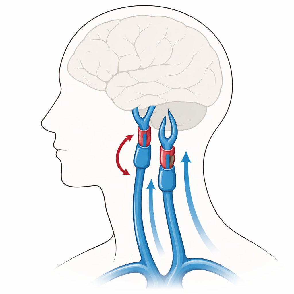
A puzzling bright spot on MRI
On arterial spin-labeling (ASL) MRI, radiologists occasionally see a patch of unusually bright signal near the left jugular bulb, a venous “junction box” where blood exits the skull into the neck. This appearance can mimic a rare but serious condition called a dural arteriovenous fistula, in which high-pressure arterial blood shunts directly into veins and can lead to bleeding, stroke, or neurological problems. Yet in many otherwise well patients, no such shunt is ever found. Earlier work suggested that mild backflow of blood up the neck vein, sometimes helped by a pinched segment near the jaw, might explain the artifact. However, many patients with this bright signal do not have that specific narrowing, hinting that other anatomical factors might be at play.
Looking closely at vein shapes
The researchers examined brain MRIs from 25 adults whose ASL scans showed this left-sided bright spot and compared them with 25 age- and sex-matched control patients without it. Both groups were chosen so that the left neck vein was relatively small compared with the right, to avoid simple dominance differences confusing the picture. Using high-resolution contrast MRI, the team carefully measured the size of the left jugular bulb at its inflow, middle, and outflow segments, as well as nearby veins that connect to it, including the posterior condylar vein that passes through a small skull channel behind the ear. They then used statistical tests and prediction models to see whether specific patterns of vein size were linked to the bright ASL signal.
Smaller outlets and side channels
Across all measurements, patients with the bright ASL signal tended to have smaller jugular bulbs and tributary veins than controls. The midportion of the jugular bulb and its outflow segment into the main neck vein were both on average narrower in the bright-signal group. Most strikingly, the posterior condylar vein was substantially thinner in these patients. When the authors corrected for multiple comparisons, only the condylar vein difference remained firmly statistically significant, but the overall pattern still suggested a consistent trend: people with the artifact tended to have a generally “slimmer” venous exit route on that side of the skull.
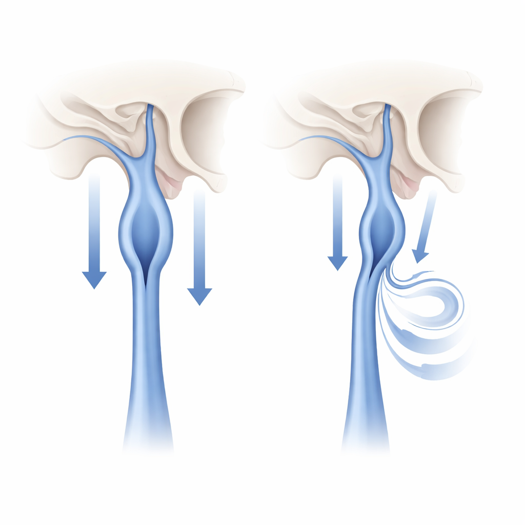
From measurements to prediction
To explore how useful these vein dimensions might be in practice, the team built models that tried to distinguish patients with the bright signal from those without it, based solely on the measured diameters. The single best predictor was the width of the posterior condylar vein, which showed moderate ability to separate the two groups. When the jugular bulb midportion, jugular bulb outflow, and posterior condylar vein sizes were combined in a multivariate model, the discrimination improved further and remained relatively stable when tested by leaving out one patient at a time. This supports the idea that it is not just one vein, but a combined anatomical configuration of relatively narrow channels, that favors slow, lingering venous blood and makes ASL more likely to “light up” that region.
What this means for patients and doctors
For now, these findings are exploratory rather than a new rulebook. The narrow posterior condylar vein approaches the limits of what current MRI scans can measure reliably, and the study was modest in size. The authors stress that their proposed threshold values should not yet be used as stand-alone diagnostic cutoffs. Still, the work offers a concrete, anatomy-based explanation for a confusing imaging finding. When a patient has a bright ASL signal at the left skull base but no clinical signs or other scan features suggesting a dangerous shunt, recognizing a pattern of relatively narrow jugular bulb segments and small side veins may help radiologists understand the signal as a benign consequence of delayed venous drainage rather than a hidden threat, guiding more measured decisions about whether invasive testing is truly needed.
Citation: Manickam, P.V., Qazi, Y., Dhawan, S.S. et al. Narrower jugular bulbs and their tributaries are associated with skull base venous hyperintensity on arterial spin-labeling MRI. Sci Rep 16, 12598 (2026). https://doi.org/10.1038/s41598-026-43549-x
Keywords: jugular bulb, arterial spin labeling MRI, brain venous drainage, dural arteriovenous fistula mimic, skull base imaging