Clear Sky Science · en
Statistical models to characterize colon tumor stiffness heterogeneity through representative atomic force microscopy maps
Feeling the Hidden Forces in Colon Cancer
When doctors look at colon tumors under a microscope, they usually focus on the shapes of cells and the presence of certain molecules. But tumors also have another, less visible trait: how stiff they are. This study explores how the "feel" of colon cancer tissue—its softness and hardness at tiny scales—relates to patient age, tumor stage, gene mutations, and other clinical factors. By combining ultra-sensitive mechanical measurements with advanced statistics and machine learning, the researchers show that tumor stiffness carries rich information that might one day help guide diagnosis and treatment.
Why Tumor Stiffness Matters
Our organs are not just bags of cells; they are woven from a supporting scaffold called the extracellular matrix, soaked in fluids and dotted with blood vessels and immune cells. This environment is not passive. It pushes, pulls, and resists, shaping how cells grow, move, and even respond to drugs. In many solid cancers, tumors tend to stiffen as collagen and other fibers build up and become more cross-linked. Cancer-associated fibroblasts, specialized support cells, are key drivers of this process. A stiffer environment can nudge cancer cells to change identity, become more invasive, and sometimes even resist therapies. In colorectal cancer, earlier work hinted that stiffness might be linked to common mutations and to how aggressive the tumor is, but a global, integrated picture was missing.
Probing Tumors with a Tiny Mechanical Finger
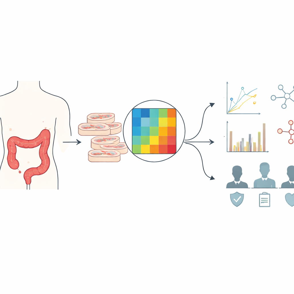
To quantify stiffness, the team used atomic force microscopy, a technique in which a microscopic spring with a rounded tip gently presses into a tissue slice. By measuring how much the tip bends for a given force, they can calculate the local stiffness at each point. For 18 patients with untreated colon cancer, they prepared thin sections from tumor and nearby healthy tissue, then recorded small square maps of stiffness, each made of a grid of indentation points covering just 50 micrometers on a side. These maps captured separate regions dominated by normal epithelium, cancer cells, fibrous stroma, or mixtures of both. After carefully filtering out noisy or incomplete measurements, the researchers analyzed 88 high-quality maps, each with dozens of reliable stiffness values.
How Healthy and Tumor Tissues Differ
The first comparison looked at healthy colon lining from areas close to the tumor and farther away. Both showed very soft values, and despite some person-to-person variation, there was no meaningful difference between these two healthy zones. Tumor tissue, however, told another story. The cancerous epithelium was clearly stiffer than normal lining, and the surrounding stroma—rich in collagen and support cells—was stiffer still. Regions where tumor and stroma mixed had intermediate stiffness, as expected from their blended composition. Sophisticated statistical models that accounted for repeated measurements within patients confirmed these trends and highlighted strong individual differences, suggesting that each person’s baseline tissue mechanics and tumor remodeling history leave a distinct signature.
From Maps to Medical Clues
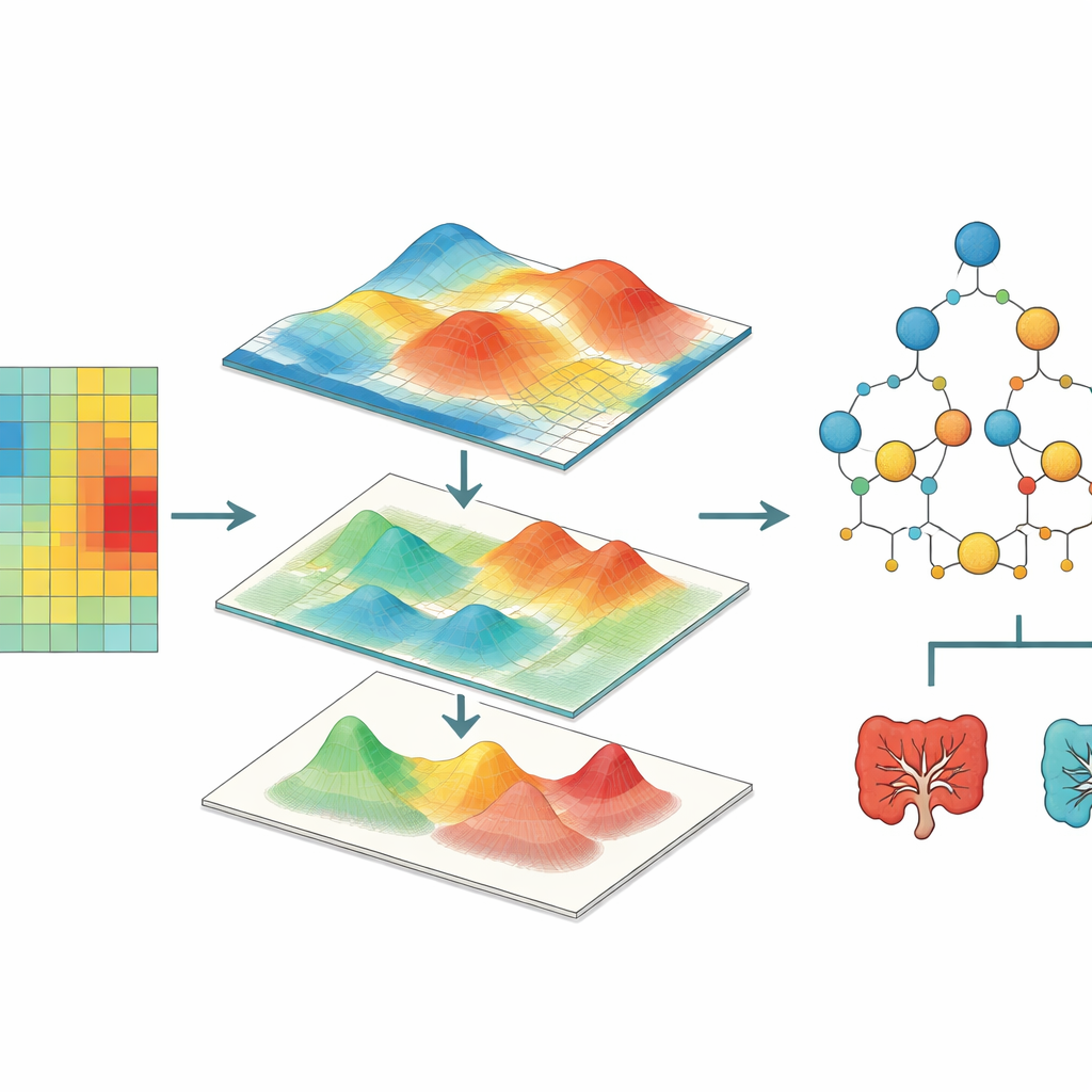
Next, the team asked whether tumor stiffness across all cancer regions taken together was linked to clinical and genetic traits. Using generalized linear mixed models, they found that stiffer patterns were associated with older age, more advanced tumor stage, and the presence of mutations in RAS genes, which are known to alter how cells sense and respond to mechanical forces. Tumors on the left and right sides of the colon, which differ in biology and patient prognosis, also showed distinct stiffness behaviors. Another notable association involved microsatellite instability, a defect in DNA repair that defines a special subtype of colorectal cancer. To go beyond average values, the researchers turned each stiffness map into a smooth surface, measured features such as roughness and patchiness, and fed these into random forest machine learning models. These models could, with moderate accuracy, infer variables like tumor stage, RAS mutation status, and whether tumor cells had invaded blood or lymph vessels.
What This Means for Patients
This work shows that the mechanical landscape of colon tumors—how stiff they are and how that stiffness varies from point to point—encodes information about tumor genetics, location, and progression. By treating stiffness maps as data-rich images and applying modern statistics and machine learning, the authors outline a framework that could eventually turn mechanical measurements into practical biomarkers. While more patients and finer maps will be needed, and the study does not yet prove direct cause-and-effect, it strengthens the idea that how a tumor feels is as important as how it looks. In the future, combining stiffness profiling with molecular tests could help doctors better classify colorectal cancers and tailor treatments to each tumor’s physical as well as genetic identity.
Citation: Gadouas, G., Tosato, G., Costa, L. et al. Statistical models to characterize colon tumor stiffness heterogeneity through representative atomic force microscopy maps. Sci Rep 16, 14314 (2026). https://doi.org/10.1038/s41598-026-43396-w
Keywords: colon cancer, tumor stiffness, atomic force microscopy, tumor microenvironment, machine learning in oncology