Clear Sky Science · en
Development and characterization of rat hindlimb ischemia models mimicking peripheral arterial disease severity
Why this matters for people with leg artery disease
Blocked arteries in the legs, known as peripheral arterial disease, can quietly rob people of their ability to walk, heal wounds, and in severe cases keep their limbs. Doctors and engineers are racing to design new treatments to grow blood vessels and protect muscle, but they first need animal models that truly mimic the different stages of this disease—from early cramping on a walk to the threat of amputation. This study builds a set of refined rat models that capture these stages in a controlled, measurable way, laying a foundation for safer and smarter testing of future therapies.
Building stepwise models of poor blood flow
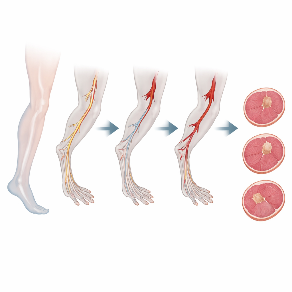
The researchers created three versions of reduced blood flow to the rat hindlimb by tying off only arteries, not veins, at different points along the main leg vessels. One model gently constricts flow high in the pelvis (the Iliac model), another cuts out a stretch of the main thigh artery (the Femoral model), and the most severe version removes segments in both the thigh and behind the knee (the Fem/Pop model). This design mirrors how leg artery disease mainly involves gradual narrowing of arteries while drainage veins remain open. It also allows scientists to dial up or down the level of injury in a consistent way, instead of relying on rough or overly destructive techniques used in many older models.
Watching blood flow and outward signs over time
To see how each operation affected circulation, the team scanned the rats’ paws repeatedly with a laser-based imaging system that measures superficial blood flow. Right after surgery, the mild Iliac model retained roughly two-thirds of its original flow, the Femoral model about half, and the Fem/Pop model less than one-third. Over the next month, flow rebounded fastest in the mild group and more slowly in the moderate and severe groups, yet by five weeks average perfusion measurements were surprisingly similar across all three. At the same time, the researchers scored visible changes in the paws—such as paleness, ulcers, and dead tissue—using a standardized scale. Here the differences were stark: the mild group quickly looked normal again, the moderate group showed temporary ulcers, and the severe group often progressed to scabs and blackened areas that only partly healed, much like advanced disease in patients.
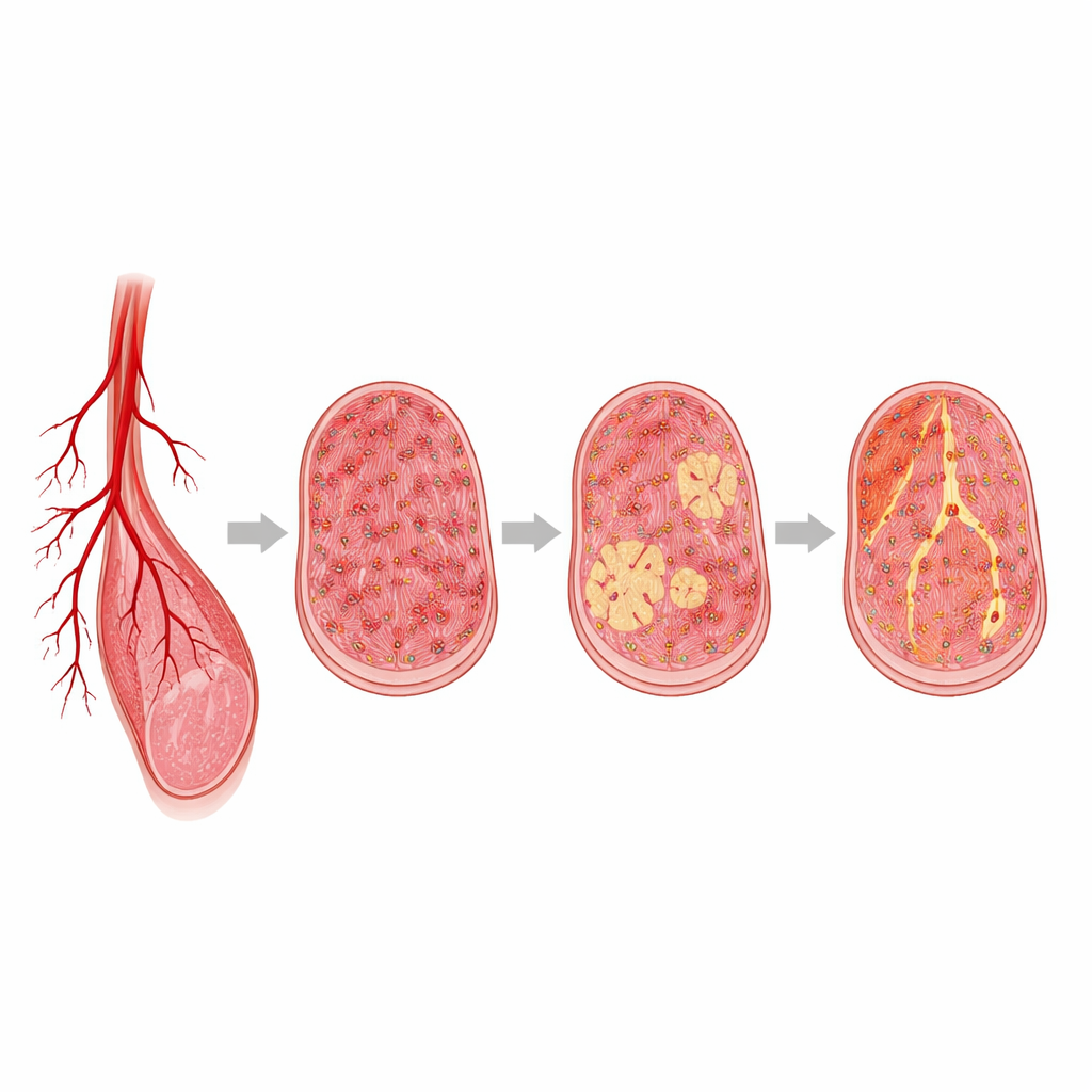
Peering inside the muscle to see lasting damage
Flow measurements alone did not tell the whole story, so the scientists examined calf muscles under the microscope after five weeks. In the mild model, muscle fibers largely kept their structure. In the moderate model, fibers thinned and showed some immune cell infiltration. In the severe Fem/Pop model, muscle architecture was heavily disrupted, with dead areas, shrunken fibers, and thick bands of scar-like tissue. The team used computer-assisted image analysis to quantify features such as fiber size and the appearance of new centrally placed nuclei, a sign that regeneration has been triggered. They found that the severe model drove both more scarring and more signs of attempted repair, while also showing the greatest loss of tiny blood vessels feeding the tissue.
Signals of stress, inflammation, and repair
Beyond structure, the study looked at the chemical and cellular environment within the injured muscles. Markers of oxidative damage—molecules formed when tissues are starved of oxygen—tended to be higher in the most severe models, although differences were modest in this small dataset. Staining for immune cells revealed that severely affected muscles harbored more of the “attacking” type of macrophages and relatively fewer of the “healing” type, suggesting a lingering inflammatory state that may block full recovery. Meanwhile, proteins that sense low oxygen and some signs of perfused microvessels persisted across all groups, indicating that even very damaged muscle retains pockets of blood flow and active signaling long after the initial injury.
A toolkit for testing tomorrow’s treatments
Taken together, these three artery-only models form a graded spectrum of leg ischemia that resembles early, middle, and late stages of human peripheral arterial disease. Rather than relying on a single readout, the framework links outward appearance, imaging-based blood flow, microscopic muscle changes, immune activity, and chemical stress into a multidimensional picture of damage and repair. The mild Iliac model is best suited to studying early interventions before major tissue loss, the Femoral model to moderate disease with partial recovery, and the Fem/Pop model to advanced limb-threatening ischemia where scarring dominates. This toolkit should help researchers more realistically judge which regenerative materials, cell or gene therapies, and engineered vessels are likely to succeed in protecting patients’ legs—and in which stage of disease they are most needed.
Citation: Liang, Y., Mullen, C., Young, E.R. et al. Development and characterization of rat hindlimb ischemia models mimicking peripheral arterial disease severity. Sci Rep 16, 12984 (2026). https://doi.org/10.1038/s41598-026-43361-7
Keywords: peripheral arterial disease, hindlimb ischemia, arterial occlusion models, muscle regeneration, angiogenesis