Clear Sky Science · en
Subchondral bone alterations in temporomandibulat joint arthralgia negatively affect the functional results of stabilization appliance therapy. A retrospective exploratory cohort study
Why Jaw Joint Pain Matters
Many people live with stubborn pain in the small joints that allow the jaw to open and close, called the temporomandibular joints. This discomfort can make simple acts like chewing, talking, or yawning difficult and exhausting. Dentists often prescribe a night‑time bite splint to ease this pain, but not everyone benefits in the same way. This study asks a practical question that matters to patients and clinicians alike: can hidden changes inside the jaw joint’s bone help predict who will improve with splint therapy and who may continue to struggle?
Looking Inside a Painful Jaw Joint
The researchers focused on people diagnosed with pain coming specifically from the jaw joint on one side, a condition known as arthralgia. They enrolled 109 adults who had persistent pain despite standard anti‑inflammatory drugs and who needed a three‑dimensional X‑ray scan called cone beam computed tomography. This scan can show small bony changes in the rounded end of the jawbone, the condyle, that ordinary X‑rays may miss. The team recorded the presence of features such as surface flattening, roughened or eroded areas, tiny bone outgrowths, scarring‑like thickening, partial loss of bone, and small cavities within the bone.
Grading Hidden Bone Damage
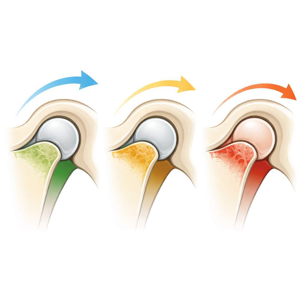
To make sense of these internal changes, the authors grouped the findings into a simple four‑step scale that reflected how far the bone seemed to have progressed along an arthritic pathway. Mild changes included just one subtle sign, while moderate changes combined several signs of wear and tear. The most advanced stages showed actual loss of bone and combinations of severe damage. Because only a limited number of patients had the very worst stage, the two highest steps were merged into a single “severe” category. This allowed the researchers to compare three practical levels: mild, moderate, and severe internal bone change beneath the joint’s smooth surface.
Testing a Common Treatment
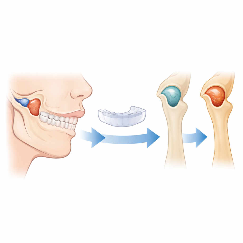
All patients received the same intervention: a resilient, three‑millimeter‑thick splint that fits over the teeth and is intended to redistribute forces in the jaw joint and reduce irritation. Before starting treatment and again after six months, patients rated their average pain over the previous week and filled out a detailed questionnaire called the Jaw Functional Limitation Scale‑20. This tool captures how much trouble they have with everyday tasks such as chewing tougher foods, opening the mouth wide, or speaking and showing emotion. By comparing scores over time, the team could see not only whether pain eased but also whether day‑to‑day function truly improved.
Who Improved the Most
Across the entire group, there was good news: average pain levels dropped sharply after six months, and all measures of jaw function improved. Yet when the researchers compared patients by their baseline bone‑damage category, important differences emerged. People whose scans showed mild subchondral bone alterations enjoyed the biggest gains in chewing ability and overall function. Those with severe underlying bone changes did improve, but their scores after six months remained noticeably worse. Moderate cases fell in between. These patterns held even after adjusting for age, sex, how long the pain had been present, and how intense it was at the start, suggesting that the hidden state of the bone itself carries unique prognostic weight.
What This Means for Patients and Clinicians
For someone living with jaw joint pain, the study’s message is twofold. First, a properly fitted splint can meaningfully reduce pain and functional limits for many people. Second, if the bone beneath the joint surface is already heavily damaged, improvement may be more modest and slower. Using three‑dimensional imaging to grade these bone changes could eventually help clinicians set more realistic expectations, decide when to complement splint therapy with other procedures, and design future studies that tailor treatment to disease stage. While more research is needed, especially to refine the grading system and account for psychological and lifestyle factors, this work suggests that what lies beneath the joint surface matters for how well a simple splint can restore comfortable, everyday jaw use.
Citation: Emshoff, R., Rudisch, A. & Bertram, S. Subchondral bone alterations in temporomandibulat joint arthralgia negatively affect the functional results of stabilization appliance therapy. A retrospective exploratory cohort study. Sci Rep 16, 13284 (2026). https://doi.org/10.1038/s41598-026-43226-z
Keywords: temporomandibular joint pain, jaw splint therapy, cone beam CT, subchondral bone changes, jaw function