Clear Sky Science · en
Spatial transcriptomic map of the mouse urinary bladder
Why Mapping a Tiny Organ Matters
The bladder is a small, easily overlooked organ—until something goes wrong. Bladder infections are common, and bladder cancer ranks among the top ten cancers worldwide. To spot early warning signs of disease, scientists first need a precise picture of what a healthy bladder looks like at the molecular level. This study delivers exactly that for mice: a detailed map showing which genes are active where inside an intact bladder, almost cell by cell. Such a reference map can help researchers recognize when and where things start to go off track in disease.
Seeing Genes Where They Live
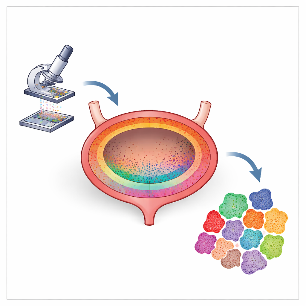
Traditional methods for studying cells often require breaking tissue apart, mixing cells into a slurry, and reading their genetic activity one by one. While powerful, this approach loses a crucial piece of information: where each cell originally sat in the organ. In this work, the researchers used a newer approach called spatial transcriptomics on thin, preserved slices of mouse bladder. Instead of scattering the cells, they laid the tissue onto a specially barcoded slide that captures messages from genes in tiny 8-by-8 micrometer squares—roughly the size of many bladder cells. After sequencing, they could reconstruct not only which genes were active, but also exactly where in the bladder wall those messages came from.
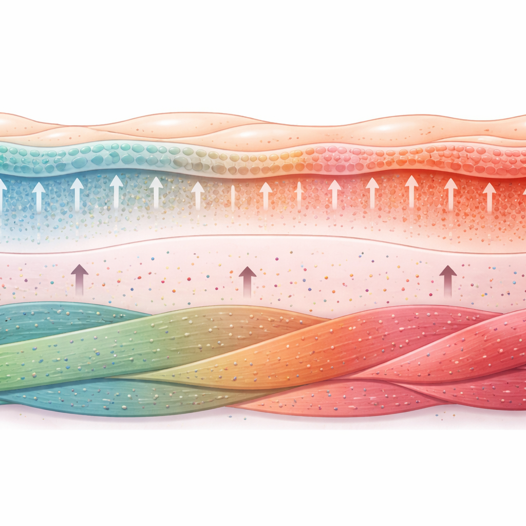
Layers of Defense Inside the Bladder Wall
The bladder wall is built like a high-tech, flexible raincoat. Its inner lining, the urothelium, must hold back acidic, waste-filled urine while stretching and relaxing many times a day. The team’s map clearly separated the three major layers: the urothelium at the surface, the supportive lamina propria beneath it, and the outer smooth muscle that powers urination. Within the urothelium, they could distinguish basal cells attached to the underlying membrane, intermediate cells above them, and large umbrella cells at the urine-facing surface—each with its own gene “signature.” Intriguingly, they also detected an additional, very topmost layer they call superficial urothelium, which may reflect subtle polarity in how umbrella cells organize their genetic activity from bottom to top.
The Hidden World Between Nerves and Muscle
Beneath the lining, the lamina propria acts as a soft cushion and communication hub. Here, the scientists found several distinct groups of fibroblasts—structural cells that build and remodel the tissue scaffold—as well as small pockets of immune cells on standby for infection or injury. Different fibroblast groups occupied different depths, matching earlier hints that not all fibroblasts are alike. Some expressed genes linked to collagen production and tissue stiffness; others carried markers linked to immune signaling or lipid handling. These patterns help explain how the bladder wall can be both strong and flexible, and how it may respond differently in various disease states, such as chronic inflammation or early tumor growth.
More Variety in Muscle Than Meets the Eye
The outer smooth muscle layer, which squeezes the bladder to expel urine, also turned out to be more diverse than previously appreciated. Earlier studies that ignored spatial context typically lumped smooth muscle cells into one broad group. Here, spatial mapping revealed four distinct smooth muscle clusters intertwined across the wall. Some clusters strongly expressed genes associated with contraction, others showed features that resemble myofibroblasts—cells that sit between muscle and fibroblasts and are important in wound healing and scarring. One cluster even combined classic muscle genes with collagen production, suggesting a role in building or maintaining the connective tissue sleeves that wrap muscle bundles. Together, these findings highlight how local environment within the wall shapes what muscle cells do.
A Reference Map for Future Disease Studies
By keeping each cell exactly where it belongs in the tissue and reading out its active genes, this study builds a high-resolution atlas of the healthy mouse bladder. It confirms known structures, uncovers new layers and cell varieties, and shows that gene activity changes gradually across the wall rather than in sharp stripes. This map gives researchers a crucial baseline: they can now compare diseased or injured bladders to see which cell types appear, disappear, or change their gene activity in specific locations. Over time, such comparisons may help explain why some infections become chronic, how scarring and stiffness develop, and which early molecular shifts foreshadow bladder cancer, ultimately guiding better diagnostics and treatments.
Citation: Matković, N., Gelemanović, A., Popović, K. et al. Spatial transcriptomic map of the mouse urinary bladder. Sci Rep 16, 13155 (2026). https://doi.org/10.1038/s41598-026-42931-z
Keywords: spatial transcriptomics, urinary bladder, urothelium, smooth muscle, tissue atlas