Clear Sky Science · en
Ultrasound-guided skeletal muscle biopsy technique permits measurement of structural, functional, cellular and biochemical properties
Why this matters for everyday health
When doctors and scientists want to understand why muscles weaken with age, disease, or injury, they often need a tiny piece of muscle to study under the microscope and in the lab. Traditional ways of taking this sample can be painful, imprecise, or yield too little tissue to answer all the important questions. This paper describes a new, ultrasound-guided muscle biopsy approach that aims to be gentler, safer, and far more informative from a single, brief outpatient procedure.
A gentler way to sample muscle
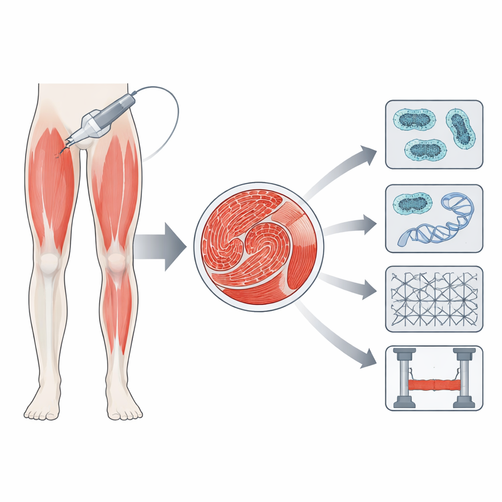
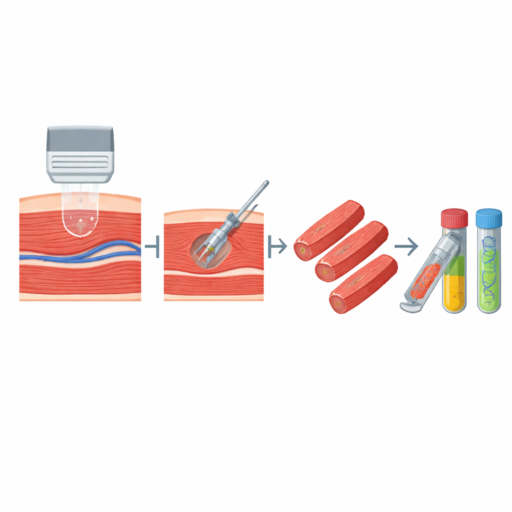
The authors combine a handheld ultrasound scanner with a battery-powered biopsy device that uses vacuum suction. Instead of several large cuts or multiple needle passes, they use a single, small skin incision and one needle insertion into two commonly studied leg muscles: the vastus lateralis in the thigh and the tibialis anterior in the shin. Ultrasound lets the operator see the muscle in real time, line up the needle along the natural direction of the fibers, and steer clear of visible blood vessels and nerves. Once the needle is in place, its internal vacuum system can draw in and cut several small “cores” of muscle without re-entering the skin.
What the team tested in volunteers
To see how well this works in people, the researchers used the technique in 19 healthy adults around 30 years old. From a single needle insertion, they routinely collected two to three samples from each muscle. The pieces were about 1.5 centimeters long and a few millimeters wide, adding up to roughly 150–170 milligrams of tissue per muscle—enough for many different tests. Immediately after the procedure, people rated their pain at about 1.5 on a 0–10 scale, and about 1.7 the next day, indicating that most experienced only mild discomfort. A few volunteers briefly felt dizzy or nauseated, and one had minor bleeding through the bandage, but all issues resolved quickly without lasting problems.
From one sample to many kinds of insight
A key strength of this method is how it turns one small biopsy into many lines of evidence about muscle health. Some tissue was frozen for classic microscopic stains that show overall structure and fiber size. Other pieces were used to measure collagen, a stiff protein that tends to build up in diseased or scarred muscle, or to examine the activity of mitochondria, the cell’s “power plants” that generate energy. The team also looked at genes involved in normal muscle function and growth, and identified muscle stem cells marked by a protein called Pax7, which are vital for repair after injury. Finally, they teased out single fibers and tested how much force they could generate and how stiff they were when stretched, giving a direct window into muscle mechanics at the cellular level.
How it stacks up against older methods
Traditional open biopsies produce large samples with well-preserved structure but require an operating room, larger incisions, sutures, and longer recovery. The classic Bergström needle and its suction-assisted versions are less invasive but can provide inconsistent sample sizes and rely on blind placement, raising the risk of hitting vessels or nerves and of collecting tissue that is less representative. Newer “microbiopsy” needles are smaller and more comfortable but often do not yield enough tissue for wide-ranging biochemical and mechanical studies. By marrying real-time ultrasound with a self-contained vacuum device that can collect multiple cores along the fiber direction, the new approach seeks a middle ground: modestly invasive, done in an outpatient setting, yet producing enough high-quality tissue for structural, mechanical, cellular, and metabolic tests all at once.
What this could mean for future patients
The authors show that this ultrasound-guided, vacuum-assisted biopsy method can reliably provide intact, well-oriented muscle samples with low pain and few short-lived side effects in healthy adults. For a layperson, the takeaway is that doctors and researchers may soon be able to learn far more about how muscles age, respond to disease, or react to new treatments, while putting patients through less discomfort and risk. Although further work is needed in older and sicker populations, and in other muscles beyond the thigh and shin, this protocol offers a promising, practical tool for studying and eventually improving muscle health.
Citation: Barber, A., Willbanks, A., Meza, G. et al. Ultrasound-guided skeletal muscle biopsy technique permits measurement of structural, functional, cellular and biochemical properties. Sci Rep 16, 12949 (2026). https://doi.org/10.1038/s41598-026-42776-6
Keywords: muscle biopsy, ultrasound-guided procedure, skeletal muscle, neuromuscular disease, muscle research