Clear Sky Science · en
Three-dimensional passive acoustic mapping of high intensity focused ultrasound fields using sparse synthetic apertures from rotated one-dimensional linear arrays
Sharper Ultrasound Aims Without Cutting
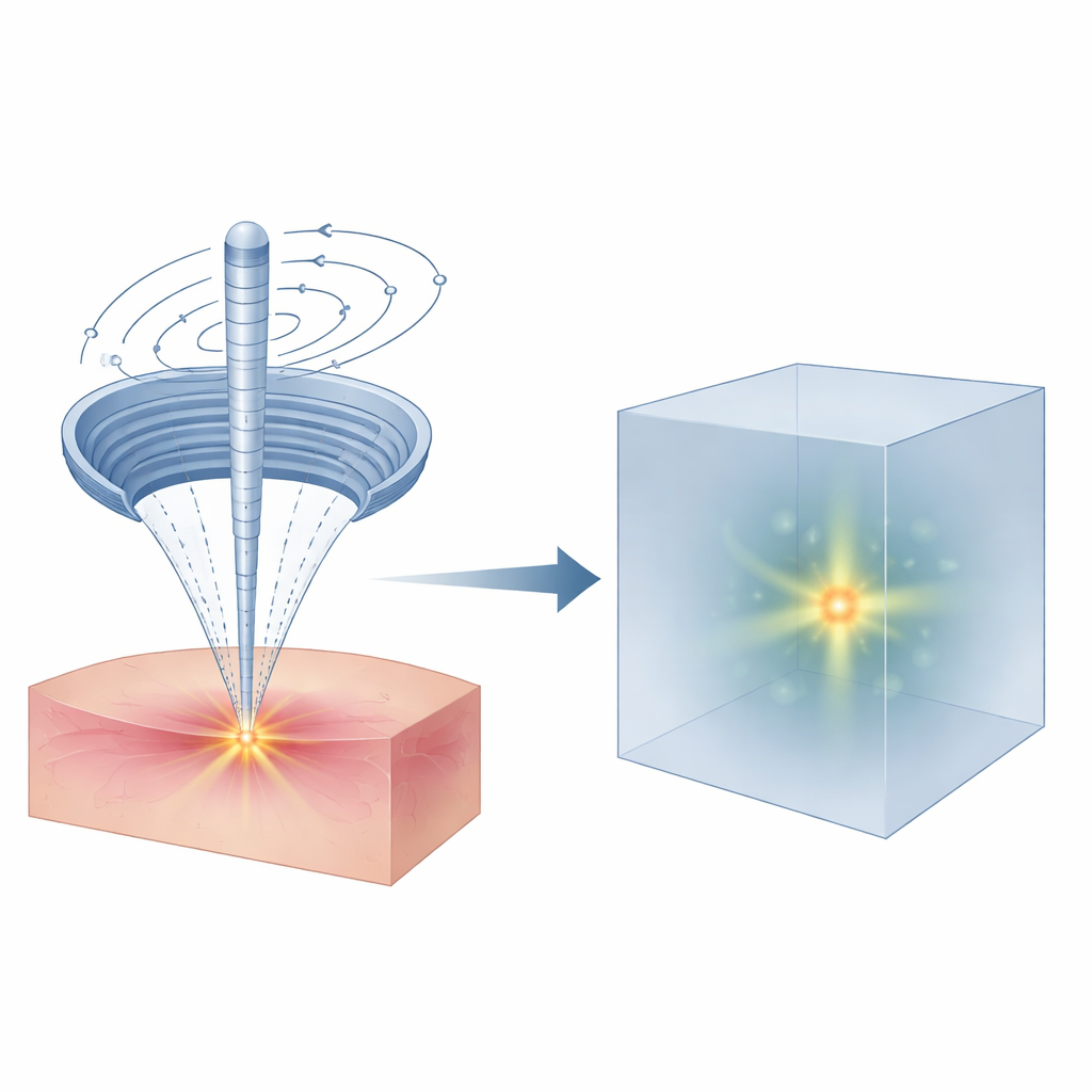
High-intensity focused ultrasound (HIFU) promises surgery without scalpels: sound waves are concentrated deep inside the body to destroy a tiny patch of tissue while leaving everything around it intact. But to be safe, doctors must know exactly where that invisible focal spot lands. This study shows how to turn faint echoes from those treatment pulses into a detailed three-dimensional map of the beam using a simple, rotating ultrasound probe and a clever spiral sampling pattern inspired by nature.
Listening Instead of Pushing
Traditional ultrasound imaging sends out sound and listens for echoes to form a picture. Here, the authors use a different tactic called passive acoustic mapping: instead of actively probing, the system “listens” to weak echoes produced when short HIFU pulses scatter off tiny variations inside tissue. By collecting these scattered signals from many angles and replaying their travel times in reverse, a computer can reconstruct where the energy was concentrated, effectively drawing a three-dimensional picture of the invisible sound field without heating or damaging tissue.
Making Do with Simpler Hardware
High-end systems that can capture full 3D sound fields usually rely on large, expensive two-dimensional arrays packed with thousands of tiny ultrasound elements. Such hardware is impractical for many therapy setups, which often must fit compactly inside other machines, such as MRI scanners. The authors turn this problem on its head: they start with a standard one-dimensional linear probe and rotate it mechanically around the HIFU beam. By choosing which probe elements to treat as “virtual” receivers at each angle, they can mimic many different two-dimensional array patterns in software, all while using only 64 channels of real hardware.
A Spiral Borrowed from Sunflowers
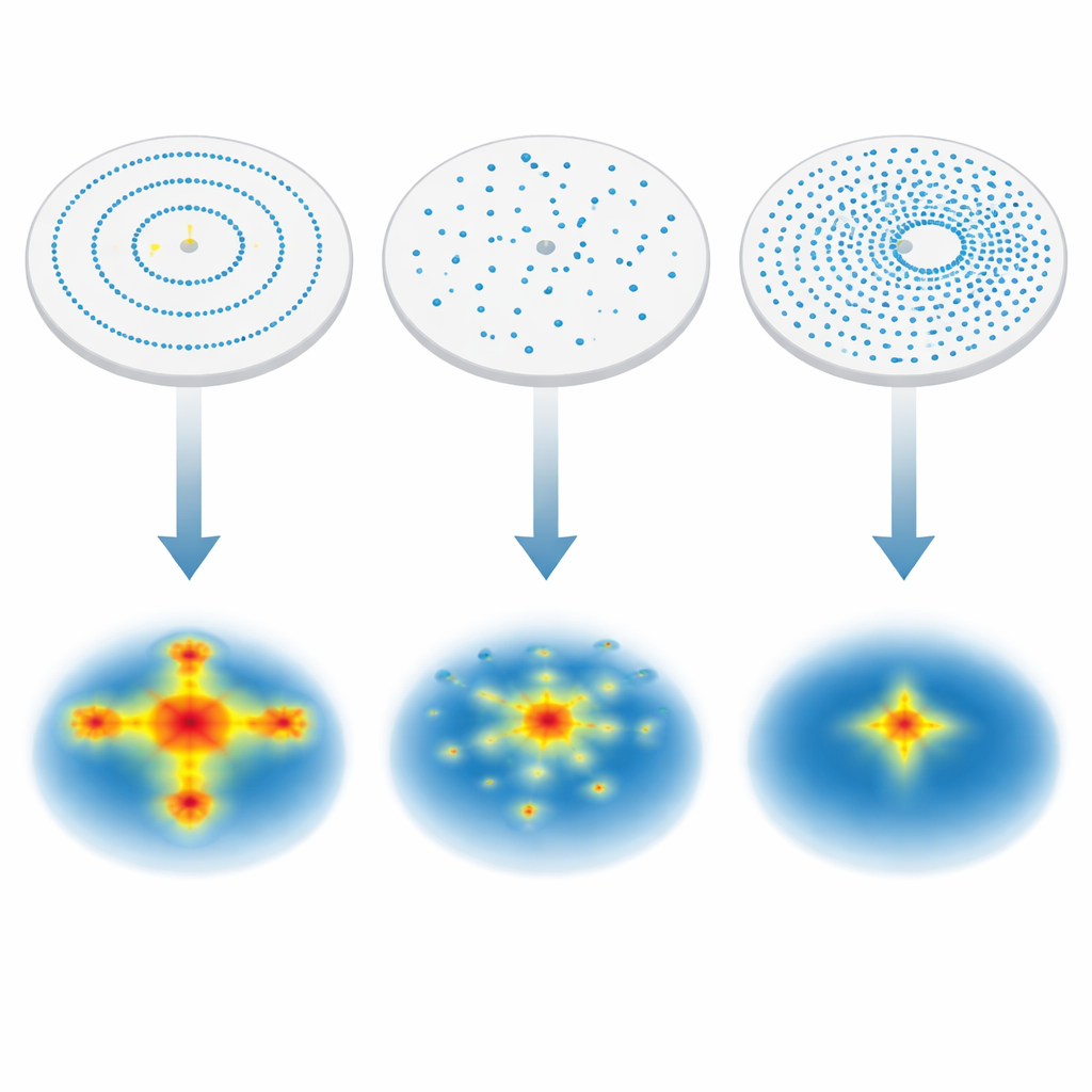
The key question is how to place—or in this case, synthesize—the receiving elements in space so that the reconstructed beam is sharp and free of misleading artifacts. The team compared six virtual layouts, including a simple straight line, a cross, concentric rings, a random arrangement, a dense full-aperture pattern, and a Fibonacci spiral that wraps elements around the circle much like seeds on a sunflower. Using detailed computer simulations of a therapeutic HIFU transducer in a tissue-like medium, they measured how faithfully each layout reproduced the true focal spot, how wide the main beam was, and how strongly unwanted sidelobes appeared.
Finding the Sweet Spot Between Order and Chaos
The results showed that layout matters as much as the number of elements. The full-aperture pattern, which uses more than 23,000 virtual channels, gave the cleanest suppression of stray energy but at the cost of heavy redundancy and data load. Highly regular rings produced a neat structure but also reinforced ring-shaped sidelobes around the focus. A purely random pattern sometimes matched the true beam well in one viewing slice, yet produced noisy halos and inconsistent performance in others. The Fibonacci spiral struck the best balance: its quasi-uniform yet nonrepetitive placement of elements yielded a compact, symmetric focal spot, accurate localization, and relatively low sidelobes in all directions, closely approaching the quality of the full-aperture reference with only a tiny fraction of the sampling.
From Simulations to Safer Treatments
In practical terms, this work suggests that a therapy system could verify where a HIFU beam will land by delivering a brief, low-energy pulse and listening with a rotating linear probe configured in a spiral sampling scheme. Within milliseconds of data collection and less than a second of mechanical motion, clinicians could obtain a three-dimensional map of the focal region before committing to stronger, tissue-ablating exposures. For patients, that means a better chance of getting the benefits of noninvasive “sound surgery” while reducing the risk of heating the wrong spot deep inside the body.
Citation: Kang, G., Hwang, J.H. Three-dimensional passive acoustic mapping of high intensity focused ultrasound fields using sparse synthetic apertures from rotated one-dimensional linear arrays. Sci Rep 16, 13711 (2026). https://doi.org/10.1038/s41598-026-42764-w
Keywords: focused ultrasound therapy, ultrasound beam mapping, passive acoustic imaging, sparse array design, Fibonacci spiral sampling