Clear Sky Science · en
Quantitative EEG signatures of power and functional connectivity alterations in Alzheimer’s disease and frontotemporal dementia
Why Brain Waves Matter for Memory Loss
Dementia touches millions of families, yet doctors still struggle to tell different forms of the disease apart early on. This study asks a simple but powerful question: can a few minutes of brainwave recordings, taken while a person rests with eyes closed, reveal reliable signs that distinguish Alzheimer’s disease from frontotemporal dementia and from healthy aging? By carefully measuring the strength and coordination of these brain waves, the researchers aim to find practical, low-cost markers that could support earlier and more accurate diagnoses.
Two Common but Different Brain Disorders
Alzheimer’s disease and frontotemporal dementia both cause gradual loss of thinking abilities, but they damage the brain in different ways. Alzheimer’s typically strikes memory-related areas and shows up first as forgetfulness in older adults. Frontotemporal dementia more often appears in middle age, altering personality, behavior, and language as frontal and temporal brain regions degenerate. Because symptoms can overlap, doctors often find it hard to decide which disease is present. The authors turned to electroencephalography (EEG)—a noninvasive method that records the brain’s electrical activity—to see whether the “rhythms” and connections of brain waves can separate these conditions from each other and from normal aging.
Listening to the Brain’s Hidden Rhythms
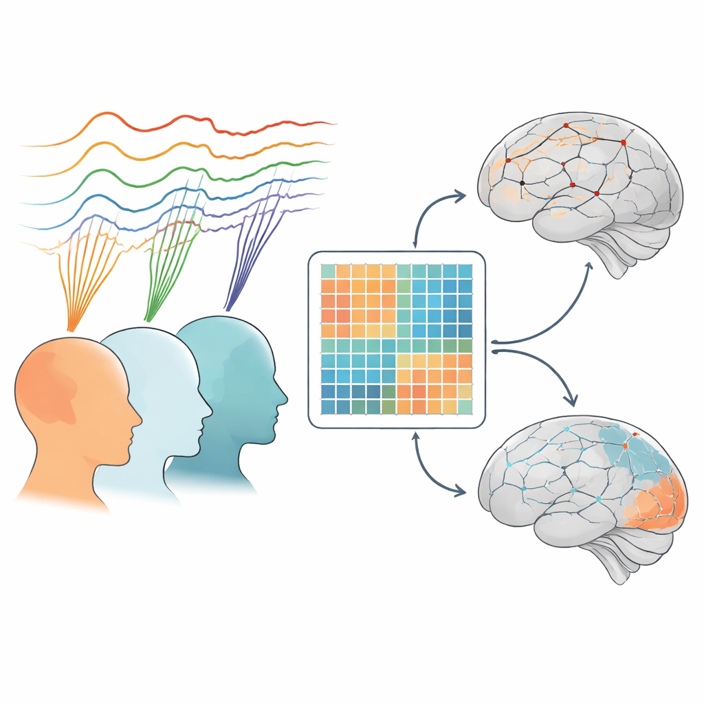
The team analyzed an open EEG dataset from 88 people: 36 with Alzheimer’s, 23 with frontotemporal dementia, and 29 cognitively normal older adults. All lay quietly with eyes closed while 19 sensors on the scalp picked up brain activity. The researchers focused on five familiar frequency bands—slow delta and theta waves, mid-range alpha, and faster beta and gamma waves. First, they measured how much power, or strength, each type of wave had across different brain regions such as frontal, temporal, parietal, and occipital lobes. Next, they examined how well these areas “talked” to each other by tracking how synchronized the waves were between pairs of sensors. Using network analysis, they summarized these connections as edge strength (individual links) and node strength (overall connectivity of each region).
Patterns of Brain Power Across the Head
One clear finding was that healthy older adults showed stronger alpha waves overall than both Alzheimer’s and frontotemporal dementia groups, especially over temporal and parietal regions in Alzheimer’s and over occipital (back-of-the-head) regions in both diseases for alpha and beta bands. In healthy brains, power was spread in a more varied way across lobes and frequencies, suggesting a rich, differentiated pattern of activity. Alzheimer’s brains showed a more uneven mix, with relatively higher slow waves and reduced fast waves in some regions, while frontotemporal dementia displayed a more uniform, flattened profile. These differences in where and how strongly particular rhythms appeared—especially in delta, theta, alpha, and gamma bands—hint that the two dementias alter brain activity in distinct spatial patterns that could help classify patients.
How Brain Regions Stay in Touch
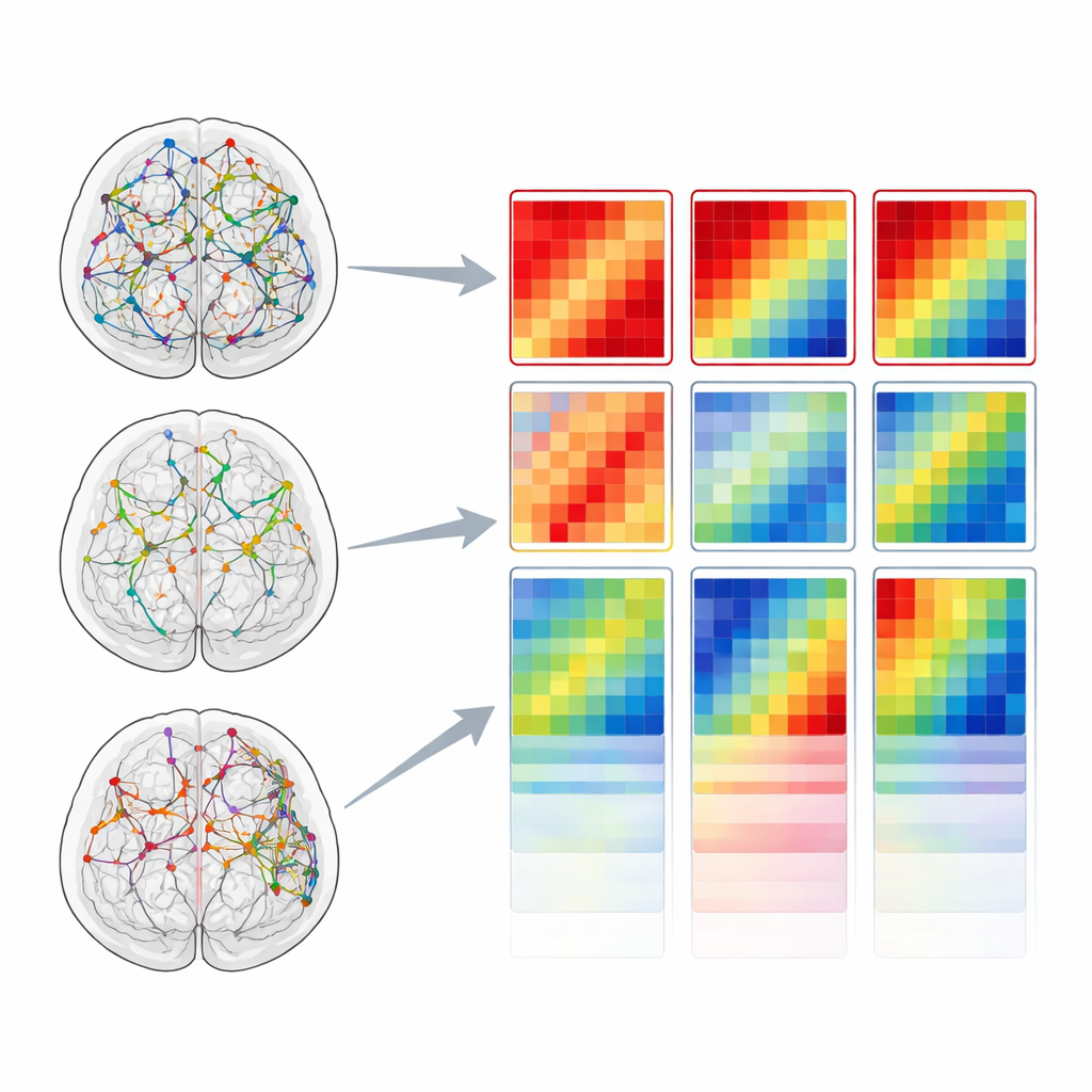
When the researchers looked at connectivity, the contrasts became even sharper. Compared with healthy adults, people with Alzheimer’s showed weaker connections between many pairs of brain areas across most frequency bands, indicating a widespread breakdown in communication. Frontotemporal dementia also showed reduced connectivity in the slow delta and theta bands, but, strikingly, displayed stronger connections in the beta band than both Alzheimer’s and control groups. Examining specific lobes, Alzheimer’s showed particularly reduced connectivity in frontal and temporal regions, while frontotemporal dementia showed its strongest disruptions in the temporal lobe but relatively spared frontal connections. Overall, frontotemporal dementia sat between normal aging and Alzheimer’s: clearly impaired, but less globally disconnected than Alzheimer’s.
What This Could Mean for Patients
Taken together, the study suggests that brief resting EEG recordings contain a double signature of dementia: changes in the strength of key brain rhythms and in the coordination between brain regions. Alzheimer’s appears as a condition with weakened alpha and beta power in certain regions and widespread network breakdown, while frontotemporal dementia shows more uniform power changes and selective, band-specific shifts in connectivity, especially in the temporal lobe. Although these results need to be confirmed in larger and more diverse samples, they point toward simple, affordable EEG measures that could help clinicians distinguish dementia types earlier, guide further testing, and ultimately support more tailored care.
Citation: Iqbal, S., Nisar, H. & Yeap, K.H. Quantitative EEG signatures of power and functional connectivity alterations in Alzheimer’s disease and frontotemporal dementia. Sci Rep 16, 12158 (2026). https://doi.org/10.1038/s41598-026-42452-9
Keywords: electroencephalography, Alzheimer’s disease, frontotemporal dementia, brain connectivity, biomarkers