Clear Sky Science · en
FMCL: a transformer-based feature-map classifier learning approach for enhanced brain tumor detection in MRI
Why spotting brain tumors sooner matters
Brain tumors are often detected using MRI scans, but even expert radiologists can struggle when tumors are small, faint, or buried in noisy images. Missing such details can delay treatment or lead to unnecessary follow‑up tests. This paper introduces a new computer‑vision method, called Feature‑Map Contrast Learning (FMCL), designed to help computers read brain MRIs more like a careful specialist—picking out suspicious regions while ignoring misleading visual clutter.
Current tools and their blind spots
Traditional computer methods for reading MRIs rely on either hand‑crafted image processing (such as filtering and edge detection) or modern deep learning models like convolutional neural networks and transformers. These approaches have made impressive progress, but they still stumble on low‑contrast tumors, images with strong noise, and differences between scanners or hospitals. Many models also need very large labeled datasets and tend to treat brightness (intensity) and texture differences (contrast) as separate or simply additive cues. In practice, that can cause one type of signal to overpower the other, making subtle tumors disappear into their surroundings or creating false alarms in healthy tissue.
A new way to balance light and texture
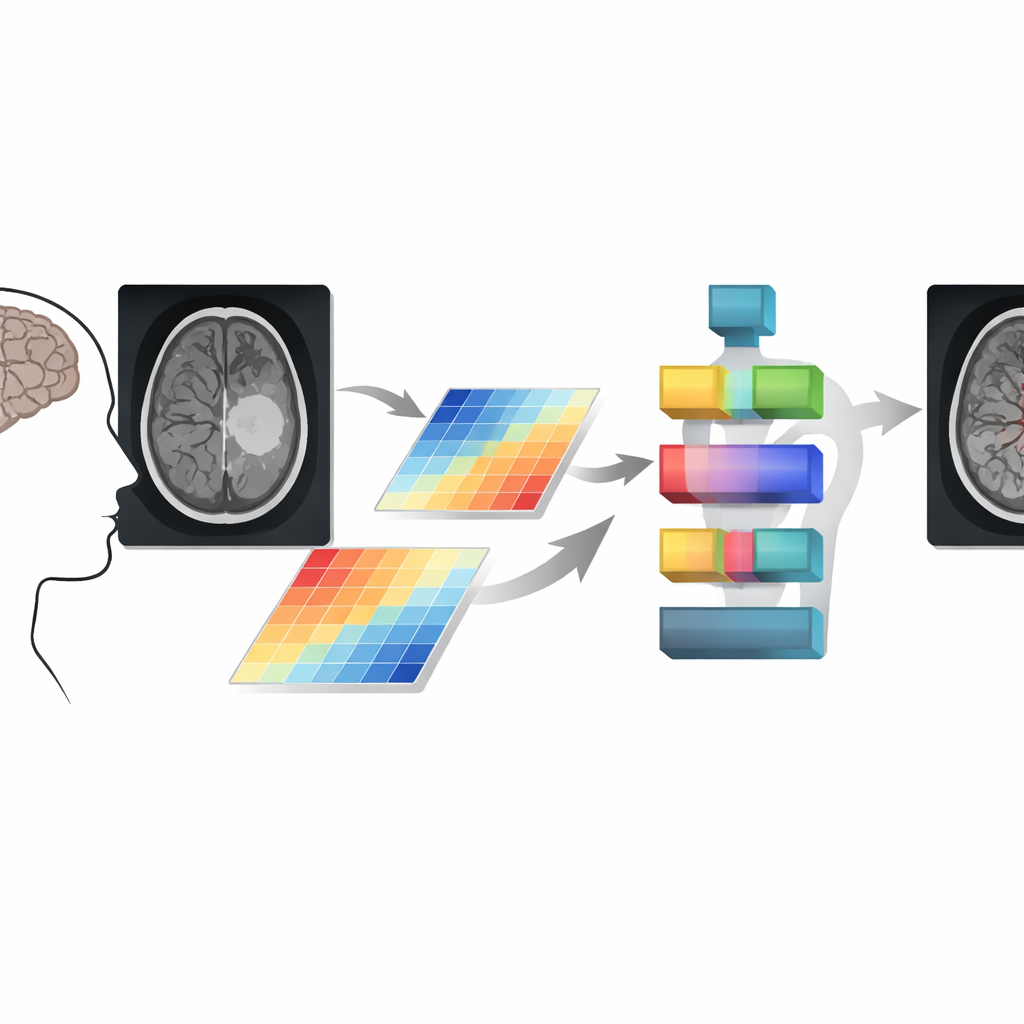
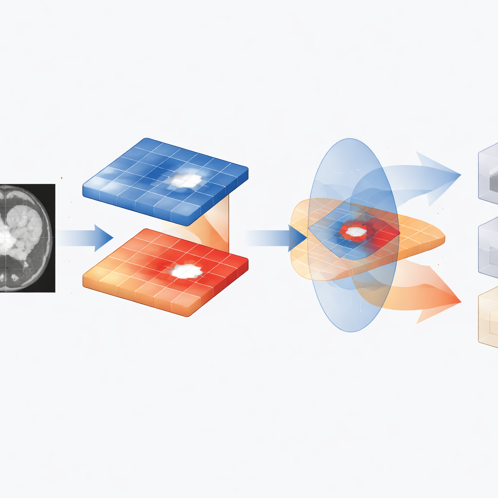
The FMCL framework tackles this by explicitly managing the relationship between contrast and intensity across every pixel in an MRI slice. The authors observe that in many real scans, bright regions often have lower contrast and vice versa; tumor tissue can flip this pattern in complex ways. FMCL builds mathematical “feature maps” that capture not only how bright or different neighboring pixels are, but also how these properties change over time or across slices. It then enforces an inverse balance between contrast and intensity so that neither dominates the decision. This careful balancing is designed to keep subtle tumors visible even when the surrounding brain tissue is unevenly lit or noisy.
Letting attention follow the tumor
Once these regulated feature maps are built, FMCL feeds them into a transformer‑based network, a type of model originally popularized in language processing and now widely used in vision. Instead of looking at raw image patches, the transformer receives already cleaned and balanced maps, and uses self‑attention to learn which regions of the brain relate to each other. The model splits regions into three main categories: clearly healthy background, clearly tumor‑affected tissue, and ambiguous low‑signal areas. It then performs a second, adjacent‑region check, where neighboring pixels are compared and smoothed using a specialized SoftMax step. This extra stage helps ensure that tumor borders are continuous and that small flickers of noise do not get mistaken for disease.
Training, testing, and performance
To evaluate FMCL, the authors used a public Kaggle dataset containing 3,264 brain MRI images labeled into four groups: no tumor, glioma, meningioma, and pituitary tumor. Images were preprocessed to standardize size, brightness, and noise before training. The model was compared with several strong deep‑learning baselines, including hybrid capsule‑and‑CNN networks and high‑capacity image classifiers. Across standard measures such as accuracy, precision, sensitivity, and recall, FMCL consistently came out ahead. It improved accuracy by about nine percentage points and reduced average classification error by roughly 11–16% compared with prior methods, while also keeping computation time per image in a range suitable for clinical use.
What this means for patients and clinics
From a lay perspective, FMCL is like giving an MRI‑reading assistant a better set of glasses and a more disciplined way of paying attention. By balancing how it weighs brightness and texture and by double‑checking boundaries between neighboring regions, the system is less likely to miss difficult tumors or over‑react to harmless variations. Although it does not replace radiologists, it can act as a second reader that flags suspicious areas and supports faster, more confident decisions. The authors note that challenges remain for very tiny or extremely faint tumors, but their results suggest that carefully designed feature‑map learning can make computer‑aided brain tumor detection more reliable and useful in real‑world hospitals.
Citation: Alanazi, T.M. FMCL: a transformer-based feature-map classifier learning approach for enhanced brain tumor detection in MRI. Sci Rep 16, 12571 (2026). https://doi.org/10.1038/s41598-026-42450-x
Keywords: brain tumor MRI, medical image analysis, deep learning, transformer models, tumor detection