Clear Sky Science · en
Single-nucleus RNA sequencing uncovers cell type-specific alterations in OSA-related liver injury
Why Nighttime Breathing Problems Matter to Your Liver
Obstructive sleep apnea, a condition in which breathing repeatedly stops and starts during sleep, is usually discussed in terms of snoring, daytime sleepiness, and heart trouble. But those brief drops in blood oxygen may also quietly injure the liver. This study used a powerful gene-reading technique to look inside thousands of individual liver cells in a rat model of sleep apnea–like oxygen dips. The goal was to see exactly which liver cells are affected and how their internal programs change, shedding light on why people with sleep apnea often show signs of fatty liver disease and liver scarring.
Breathing Pauses and Oxygen Swings
In people with obstructive sleep apnea, the throat repeatedly narrows or collapses during sleep, causing episodes of low oxygen followed by re‑oxygenation. To mimic this pattern, the researchers exposed rats to cycles of normal and low oxygen for eight hours a day over twelve weeks, while a comparison group breathed normal air. When the livers were examined under the microscope, the oxygen‑stressed animals showed clear injury: scattered patches of dead liver cells and heavy clusters of inflammatory cells, especially around the vessels that bring blood into the liver. These changes resemble early stages of liver inflammation and damage reported in people with sleep apnea and non‑alcoholic fatty liver disease. 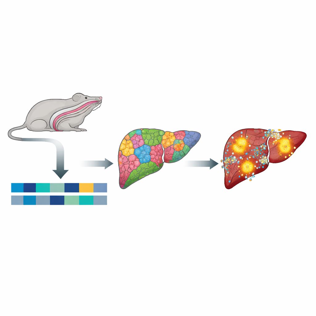
Reading the Messages Inside Individual Liver Cells
Instead of grinding the liver into a single mixed sample, the team isolated nuclei from individual cells and sequenced their RNA—essentially reading which genes were turned on or off in each cell. From over 70,000 nuclei, they identified ten major liver cell types, including the main workhorse liver cells (hepatocytes), blood vessel lining cells, supporting stellate cells, and several kinds of immune cells such as macrophages and T cells. Interestingly, the overall proportions of these cell types did not change much between normal and oxygen‑stressed animals. What did change was the internal activity of the cells: their gene expression patterns were extensively rewired, revealing a liver that looks similar in composition but behaves very differently at the molecular level.
Energy Use, Fat Handling, and Scarring Signals Go Off-Kilter
Hepatocytes and stellate cells showed some of the strongest shifts. Genes involved in burning fats and maintaining healthy metabolism were dialed down, including pathways controlled by PPAR, a key regulator that helps the liver process fatty acids and avoid fat buildup. At the same time, stress‑response and survival pathways such as AMPK and PI3K–Akt were turned up, suggesting that cells were trying to adapt to the repeated oxygen swings. In stellate cells—normally quiet helpers that support the liver’s structure—genes linked to cell adhesion and collagen production became more active, pointing toward a shift toward scar‑forming behavior. Together, these changes sketch a picture of a liver moving away from balanced fat handling and toward inflammation and fibrosis.
Blood Vessels and Immune Cells Join the Disturbance
The cells that patrol and line the liver’s blood channels were also reshaped. Liver macrophages, the resident “clean‑up” immune cells, switched on gene programs tied to inflammation and blood clotting, including the NF‑κB pathway and complement and coagulation cascades, which can drive chronic injury and scarring. Endothelial cells, which form the inner lining of liver vessels, altered genes that control their internal skeleton and tight junctions, changes that can weaken the barrier between blood and tissue and affect how immune cells move into the liver. T cells showed shifts in genes related to metabolism and stress within the protein‑folding machinery, hinting that their behavior and staying power in the liver microenvironment are also changed by intermittent low oxygen. 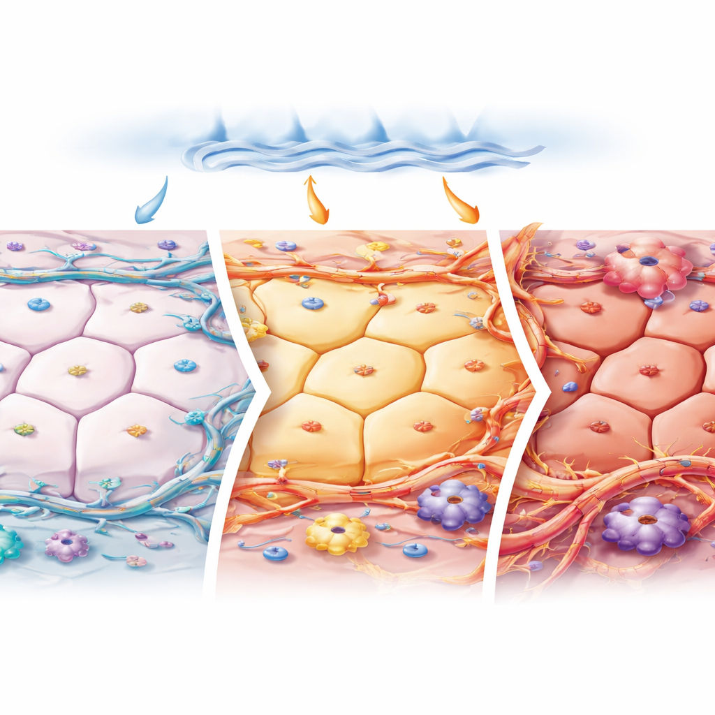
What This Means for People with Sleep Apnea
By mapping how each type of liver cell responds to repeated drops in oxygen, this study provides a detailed atlas of early liver injury in a sleep apnea–like setting. Rather than simply killing cells outright, intermittent hypoxia appears to reprogram many liver and immune cells at once—reducing healthy fat processing, activating inflammatory and clotting pathways, and nudging support cells toward scar formation. Although this work was done in rats and focuses on gene activity rather than symptoms in patients, it helps explain why sleep apnea is so strongly linked to fatty liver disease and fibrosis. It also highlights potential targets—such as pathways controlling fat metabolism, inflammation, and stellate cell activation—that future therapies might aim to calm in order to protect the livers of people living with obstructive sleep apnea.
Citation: Huang, WS., Wang, CQ., Huang, YZ. et al. Single-nucleus RNA sequencing uncovers cell type-specific alterations in OSA-related liver injury. Sci Rep 16, 11522 (2026). https://doi.org/10.1038/s41598-026-42236-1
Keywords: obstructive sleep apnea, liver injury, intermittent hypoxia, single-nucleus RNA sequencing, fatty liver disease