Clear Sky Science · en
Human stem cell-derived neurons establish functional inhibitory–excitatory cortical circuits in a chimeric transplantation model
Why balancing brain signals matters
Our thoughts, movements and memories depend on a delicate balance between brain cells that excite activity and those that calm it down. When this balance tips too far in either direction, problems such as epilepsy, autism, stroke damage or neurodegenerative diseases can emerge. This study explores whether human stem-cell–derived brain cells can be transplanted into a mouse brain in a way that rebuilds both sides of this balance—excitatory and inhibitory neurons—so that they form working circuits together.
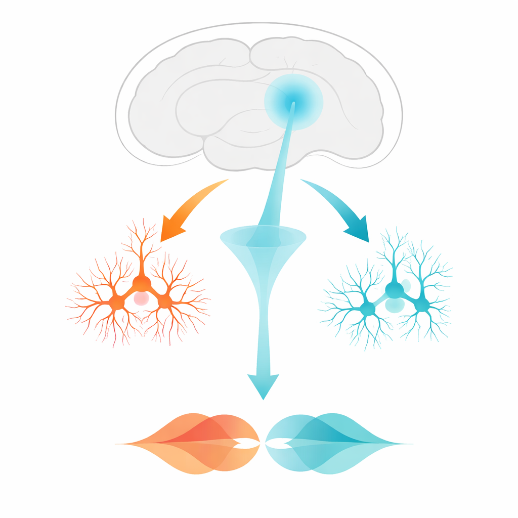
Two types of brain cells from human stem cells
The researchers started with human embryonic stem cells and guided them along two distinct developmental paths. One path produced excitatory cortical neurons, the kind of cells that send “go” signals and normally make up most of the outer layer of the brain. The other path produced inhibitory interneurons from a region called the medial ganglionic eminence, cells that act more like brakes and fine-tune the activity of their neighbors. Using fluorescent tags, the team could visually distinguish these two human cell types and track them over time. Laboratory tests showed that each group adopted the expected molecular markers and shapes of their target cell type.
Building a mixed human circuit in the mouse brain
To test whether these two human cell populations could live and work together in a real brain, the scientists transplanted a mixture of excitatory and inhibitory neurons into the cortex of adult mice. They then waited ten months—long enough for human neurons to mature. When they later examined the brains, the transplanted cells had survived, spread fibers into surrounding mouse brain regions, and developed into the expected excitatory and inhibitory subtypes. Although the final proportion of inhibitory cells was higher than what is normally found in cortex, both groups of human neurons formed dense networks with one another and with nearby host tissue.
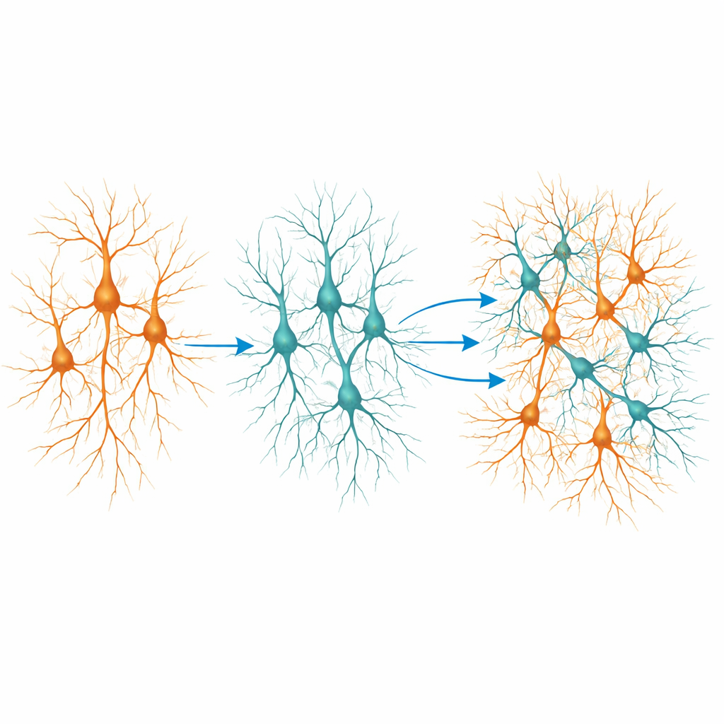
Turning cells on with light to test the wiring
Showing that cells sit in the right place is not enough; they must also communicate properly. To probe this, the team engineered the inhibitory human neurons to carry a light-sensitive protein. With this tool, shining blue light onto the graft could selectively activate the inhibitory cells. Using fine-tipped electrodes in brain slices, the researchers recorded electrical signals from both excitatory and inhibitory human neurons inside the graft. They found that the transplanted neurons displayed mature electrical properties, receiving spontaneous inputs from the surrounding network. Crucially, when the inhibitory neurons were activated by light, many excitatory human neurons showed characteristic inhibitory signals—brief drops in voltage that reflect “slow down” messages.
Proof that the brake cells really work
To confirm that these signals were genuinely inhibitory, the researchers added a drug that blocks GABA, the main chemical used by inhibitory neurons. Under this blockade, the light-triggered inhibitory responses in excitatory cells disappeared, showing that the signals were indeed carried by the transplanted inhibitory neurons using their natural messenger. Some excitatory-like responses were also observed, likely due to a small fraction of cells that did not follow the intended developmental path, but the predominant effect was inhibitory. Together, these experiments demonstrate that human stem-cell–derived inhibitory interneurons can form functional connections onto human excitatory neurons after transplantation and actively shape their activity.
What this could mean for future brain repair
This work shows that it is possible to rebuild not just isolated neurons but working microcircuits that include both accelerator and brake cells in the brain. For conditions like stroke, where large swaths of cortex are lost, such chimeric grafts may one day offer a way to restore more natural patterns of activity rather than simply adding extra excitation. The same approach could be used to study diseases where excitation–inhibition balance is disturbed, by creating long-lived human neural networks in animals from patient-derived cells. While many hurdles remain—such as refining cell ratios, cell types and safety—this study provides a key proof-of-principle that complex human cortical circuits with built-in inhibitory control can be reconstructed in the living brain.
Citation: Hunt, C.P.J., Thek, K.R., Durnall, J. et al. Human stem cell-derived neurons establish functional inhibitory–excitatory cortical circuits in a chimeric transplantation model. Sci Rep 16, 12144 (2026). https://doi.org/10.1038/s41598-026-42112-y
Keywords: stem cell transplantation, cortical circuits, excitation inhibition balance, inhibitory interneurons, stroke therapy