Clear Sky Science · en
Postmenstrual age-adjusted brain volumes and their association with early neurodevelopmental outcomes in very low birth weight infants
Why tiny brains matter for a lifetime
Every year, more babies survive after being born very early and very small, but many of them face learning, language, or movement challenges as they grow. Parents and doctors urgently want to know which infants are at highest risk so that therapy and support can start as soon as possible. This study explores whether routine brain scans near the time a premature baby would have been full-term can offer a simple, practical way to forecast early development—especially by looking at how big certain parts of the brain have grown.
Babies born too soon and their hidden risks
Very low birth weight infants—those weighing less than about one kilogram at birth—are far more likely than full-term babies to experience delays in thinking, talking, and moving. These problems can appear even when standard brain imaging does not show obvious damage. Although bedside ultrasound is widely used, it cannot capture the brain’s fine structure as well as magnetic resonance imaging (MRI). As MRI has become more available in newborn units, researchers have begun to ask not just whether the brain looks injured, but how its size and growth might predict a child’s future abilities.
Measuring growing brains with simple numbers
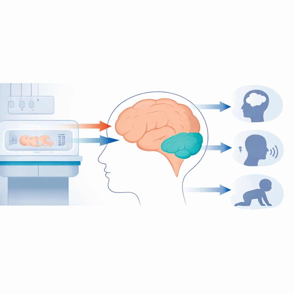
In this study from a large neonatal intensive care unit in Korea, 118 very low birth weight infants had MRI scans around the time they reached the equivalent of 40 weeks of pregnancy. Using specialized but semi-automatic software, technicians measured the overall brain, the large upper part (the cerebrum), the smaller lower part at the back (the cerebellum), the brain stem, and the fluid-filled spaces. Because scans were done at slightly different ages, the team mathematically adjusted each measurement to what it would be at exactly 40 weeks of postmenstrual age, then converted those values into z-scores—standardized numbers showing whether a baby’s brain was larger or smaller than average for that age.
Linking brain size to early development
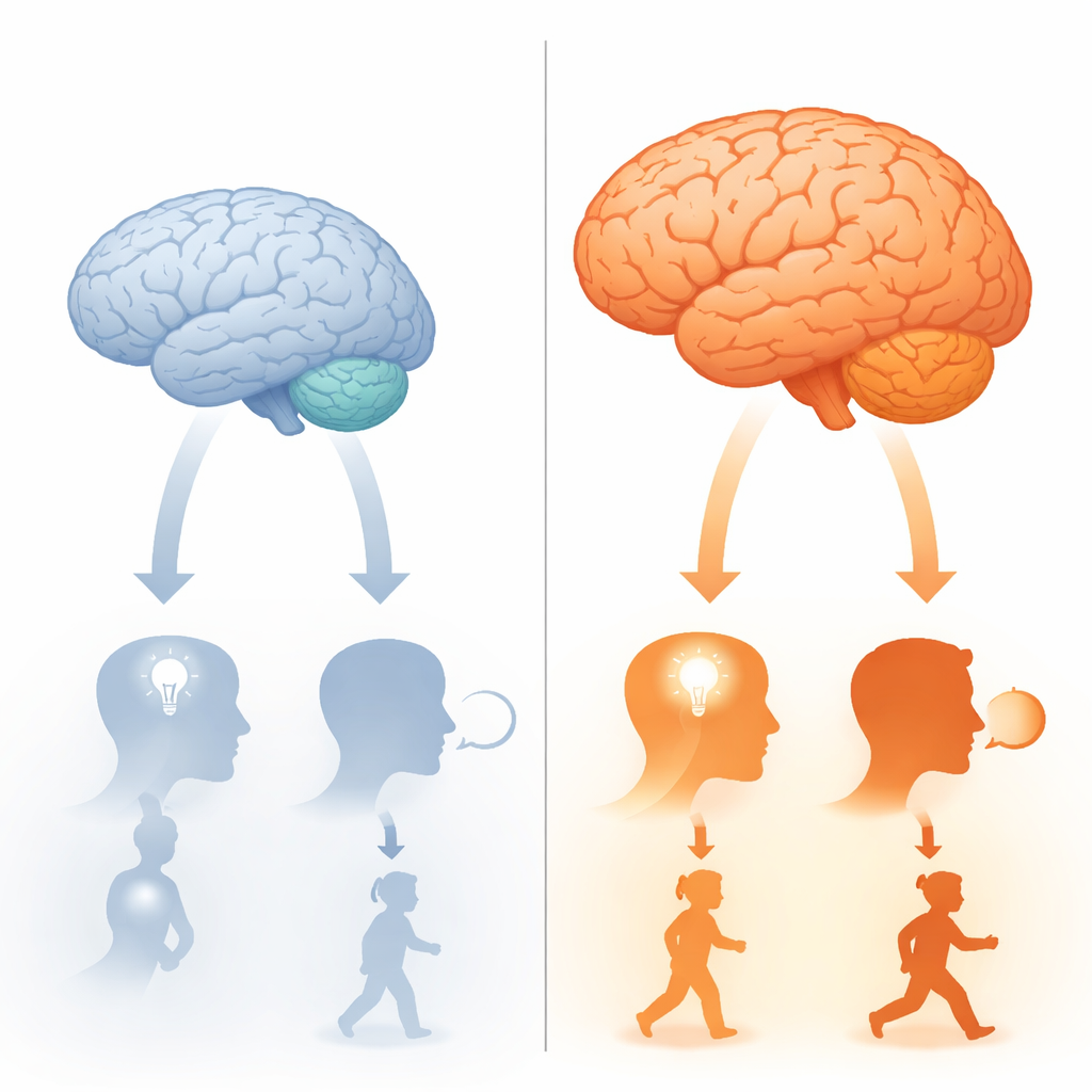
Seventy-five of these infants later completed a detailed developmental test at 12 to 18 months corrected age, assessing thinking, language, and motor skills. When the researchers compared brain size with these scores, one region stood out: the cerebellum. Babies with larger cerebellar volumes tended to have better performance across all three areas, even after accounting for sex. Total brain size and cerebrum size were also related to language and motor skills, but their links were a bit weaker. When infants were divided into “smaller” and “larger” brain groups, those with smaller cerebellums had notably lower language scores, suggesting that undergrowth in this region may be an early warning sign for later speech and communication problems.
What shapes these small brains
The study also showed that babies with smaller brains had arrived earlier in pregnancy, weighed less at birth, and had more medical complications such as lung disease, infections, and brain-related problems like white matter injury or bleeding. By the time they left the hospital, these infants were lighter, shorter, and had smaller head sizes than peers with larger brains, and they stayed in intensive care longer. Together, these findings suggest that both the stress of being born too soon and the struggles that follow in the neonatal unit can slow brain growth, which in turn may influence how a child learns and moves in the first years of life.
Turning scans into early action
For families and clinicians, the key message is that a relatively straightforward way of reading newborn MRIs—measuring the size of major brain regions and adjusting for age—can offer meaningful clues about which very small infants may face early developmental challenges. In particular, a smaller cerebellum near term-equivalent age appears to flag increased risk for language delay. While the study was modest in size and focused on outcomes only up to 18 months, it supports the idea that simple, age-adjusted brain volume measures could complement traditional imaging to guide follow-up and early intervention. With future advances, including automated and AI-based tools, such measurements might become a routine part of caring for the tiniest and most vulnerable newborns.
Citation: Oh, MY., Kim, S., Kim, M.S. et al. Postmenstrual age-adjusted brain volumes and their association with early neurodevelopmental outcomes in very low birth weight infants. Sci Rep 16, 12921 (2026). https://doi.org/10.1038/s41598-026-42039-4
Keywords: preterm infants, very low birth weight, brain MRI, cerebellar development, early neurodevelopment