Clear Sky Science · en
Upper limb lymphatic mapping and quantitative functional analysis in normal cynomolgus monkeys using indocyanine green near-infrared fluorescence lymphography
Why the Hidden Plumbing of the Arm Matters
Our arms contain a quiet but vital plumbing system that helps defend us from infection and keep swelling in check: the lymphatic network. When this system is damaged—often after breast cancer surgery—fluid can build up, leading to chronic, sometimes debilitating arm swelling called lymphedema. To prevent and better treat this condition, researchers need a clear picture of how a healthy arm’s lymphatic system is arranged and how strongly it pumps. This study uses advanced fluorescent imaging in monkeys closely related to humans to map that hidden network and measure how it works in real time.
Following the Flow from Hand to Armpit
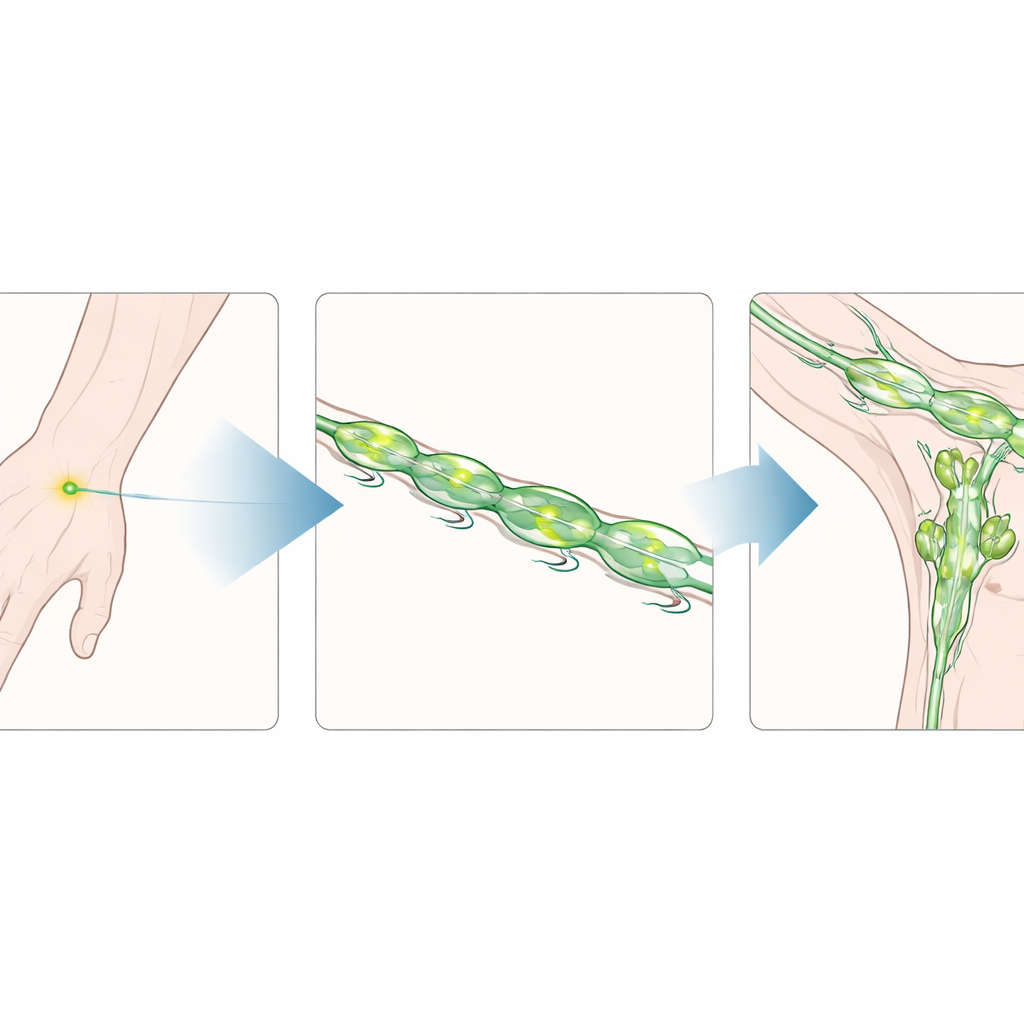
The researchers worked with five healthy cynomolgus monkeys, a nonhuman primate species frequently used in medical research. They injected a harmless fluorescent dye just under the skin between the fingers and on the palm, then used near‑infrared cameras to watch the dye move through the arm’s lymphatic vessels. These are the small tubes that carry immune cells and excess fluid toward filters called lymph nodes. The team focused on the superficial vessels just beneath the skin, tracking how they run from the hand up toward the armpit region, where a key cluster of nodes, the axillary lymph node basin, sits.
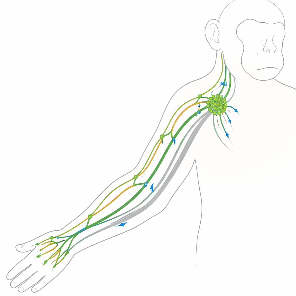
One Main Drainage Basin for the Arm
Across all ten arms studied, a strikingly consistent pattern emerged. Lymphatic vessels from the back of the hand followed paths that paralleled two familiar veins, known in people as the cephalic and basilic veins, and then all converged toward the axillary nodes in the armpit. Fluid from the palm did not take a separate shortcut; instead, it merged into the same dorsal pathways in the forearm before heading upward. Almost no superficial drainage appeared along the outer side of the upper arm. These findings suggest that, at least in these monkeys, the superficial lymphatics of the arm behave like a single drainage basin that funnels fluid into one main outlet in the armpit.
Why a Single Pathway Increases Risk
This single‑basin layout has important implications. In the leg, earlier work in the same species showed two major superficial drainage regions, offering some redundancy if one path is blocked. In the upper limb, by contrast, having most superficial flow depend on a shared route into the axillary nodes could make the system more vulnerable. If these armpit‑bound pathways are damaged during surgery or radiation, there may be fewer alternative routes for fluid to escape, raising the likelihood of chronic swelling. Although some small side paths may have been missed due to the chosen injection sites, the dominance of this common route helps explain why arm lymphedema is such a frequent complication of breast cancer treatment.
Watching the Lymphatic Pump in Action
The study went beyond anatomy to assess how vigorously the lymphatic vessels pump. By analyzing brightness changes of the fluorescent signal over time in selected regions of the arm, the team could see the rhythmic surges that mark each contraction of the vessel wall. They combined a traditional method that counts peaks in the signal with a more sophisticated time‑frequency analysis that can handle irregular, non‑clocklike rhythms. In these healthy monkeys, the pumping frequency and strength were measurable and reasonably consistent between animals, even though the overall travel time of dye from hand to elbow or armpit varied quite a bit from one individual to another. Importantly, the key pumping metrics stayed stable over the first 15 minutes after dye injection, showing that researchers can sample during this window without worrying much about timing.
What This Means for Future Patients
By carefully mapping where fluid travels in a healthy primate arm and how forcefully the lymphatic vessels contract, this work builds a baseline reference for future disease studies. Because cynomolgus monkeys share many anatomical and physiological traits with humans, these findings help bridge the gap between rodent experiments and clinical observations in people. In practical terms, the study shows that near‑infrared fluorescence imaging can noninvasively capture both the layout and the pumping behavior of arm lymphatics, and that these measurements are stable enough to be useful for comparison. As researchers begin to model lymphedema and test new treatments in primates, this "normal map" of drainage pathways and pumping patterns will serve as a crucial yardstick for detecting when, where, and how the system breaks down.
Citation: Yang, J., Jeon, E., Kim, J. et al. Upper limb lymphatic mapping and quantitative functional analysis in normal cynomolgus monkeys using indocyanine green near-infrared fluorescence lymphography. Sci Rep 16, 13090 (2026). https://doi.org/10.1038/s41598-026-42008-x
Keywords: lymphedema, lymphatic imaging, indocyanine green, nonhuman primate model, upper limb lymphatics