Clear Sky Science · en
ResNet based backbone integrated YOLO framework for bone fracture detection
Smarter Help for Broken Bones
When someone shows up in the emergency room with a possible broken wrist, minutes matter—and so does accuracy. Yet tiny cracks in children’s bones can be surprisingly hard to spot on grainy X-ray images, even for experienced specialists. This paper presents a new artificial intelligence (AI) system, called ResYOLO11, designed to act as a fast, careful second set of eyes on wrist X-rays, helping doctors find fractures more reliably while keeping computer demands low enough for everyday hospital use.
Why Reading X-Rays Is So Hard
Bone fractures are among the most common injuries worldwide, especially for children and older adults. On wrist X-rays, the telltale lines and subtle bends that signal a break can be faint and obscured by noise, low contrast, or overlapping structures. Traditional computer methods, which rely on simple edges and brightness patterns, often miss these details. Even modern deep-learning systems that excel on everyday photographs can struggle, because medical images look very different from the colorful, high-contrast pictures those systems were originally built for.
Blending Two Powerful Ideas
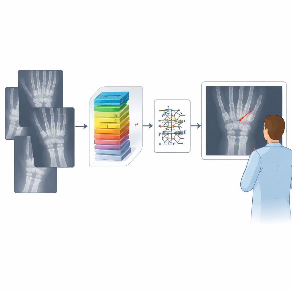
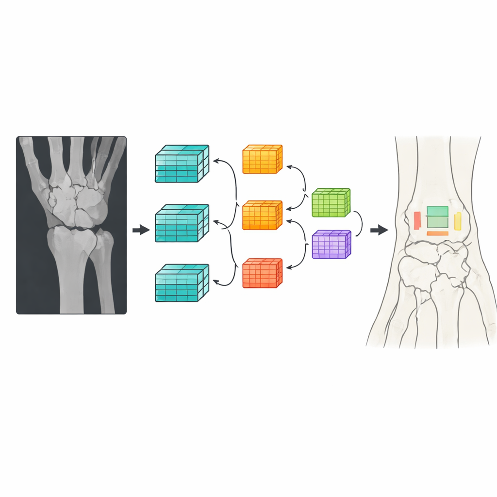
The authors tackle this challenge by combining two influential AI concepts into one unified model. One part, known as ResNet, is especially good at teasing out fine structural details from images by using “skip connections” that let information flow around layers instead of fading away. The other part, a family of detectors known as YOLO, is built for extremely fast object detection—drawing boxes around things of interest in a single pass. ResYOLO11 uses a customized ResNet50 section as its backbone to extract rich features from X-ray images, then passes them into a streamlined YOLO11 detection head that quickly proposes likely fracture locations and other abnormalities in one stage.
Cleaning Up the Picture Before the Machine Looks
Because raw X-rays can be noisy and low contrast, the team designed a careful image-preparation pipeline before the AI ever sees an image. Every wrist X-ray is resized to a standard square format and then passed through several filtering steps that smooth away speckled noise while preserving sharp bone edges. Finally, a contrast enhancement method brightens subtle boundaries so that hairline fractures stand out more clearly. Through controlled experiments, the researchers show that each extra enhancement step provides a small but consistent boost in detection quality, and that a medium image size offers the best trade-off between accuracy and speed for real-world clinical use.
How Well the New System Performs
To test their approach, the authors trained and evaluated ResYOLO11 on a large public dataset of pediatric wrist X-rays that includes over ten thousand studies and detailed expert annotations. They compared several sizes of their model—ranging from a compact “nano” version to an extra-large one—against corresponding standard YOLO11 models. Across all sizes, ResYOLO11 was more precise at identifying fractures, meaning it raised fewer false alarms, and achieved higher overall detection scores that measure both whether a fracture is found and how well its location is outlined. For the largest version, the model correctly identified fractures with very high accuracy while also cutting model size roughly in half and speeding up predictions by about a quarter compared with the baseline. Tests on a separate dataset of fractures from different body parts showed that the method generalizes beyond pediatric wrists.
What This Could Mean for Patients
The authors emphasize that ResYOLO11 is intended to support, not replace, orthopedic surgeons and radiologists. In busy emergency rooms or clinics with limited specialists, such a tool could quickly highlight suspicious regions on an X-ray, helping doctors focus their attention and reducing the chance that a subtle break is overlooked. At the same time, its relatively small memory footprint and fast response make it realistic to run on standard clinical hardware. With further validation on more age groups and body regions, and with added tools to explain its decisions, systems like ResYOLO11 could become reliable partners in the exam room, quietly checking every image to make sure no broken bone goes unnoticed.
Citation: Bhattacharya, D., Das, S., Biswas, T. et al. ResNet based backbone integrated YOLO framework for bone fracture detection. Sci Rep 16, 12954 (2026). https://doi.org/10.1038/s41598-026-41782-y
Keywords: bone fracture detection, medical imaging AI, X-ray analysis, deep learning, object detection