Clear Sky Science · en
Facial landmark-based localization of the parotid duct: a cadaveric study with ultrasonographic feasibility assessment
Why the Cheek’s Hidden Tube Matters
Inside each of our cheeks runs a tiny tube that we almost never think about: the parotid duct, which carries saliva from a large gland near the ear into the mouth. Because this duct lies close to the skin, it is easily bumped, cut, or squeezed during cosmetic procedures, facial surgery, or after injuries. Damage can lead to pain, swelling, or leaks of saliva under the skin. This study set out to map exactly where that duct usually lies on the face, using everyday features like the ear, lips, and eyes as guideposts, and to check whether modern ultrasound can see it in living people using those same landmarks.
Following the Saliva Pathway
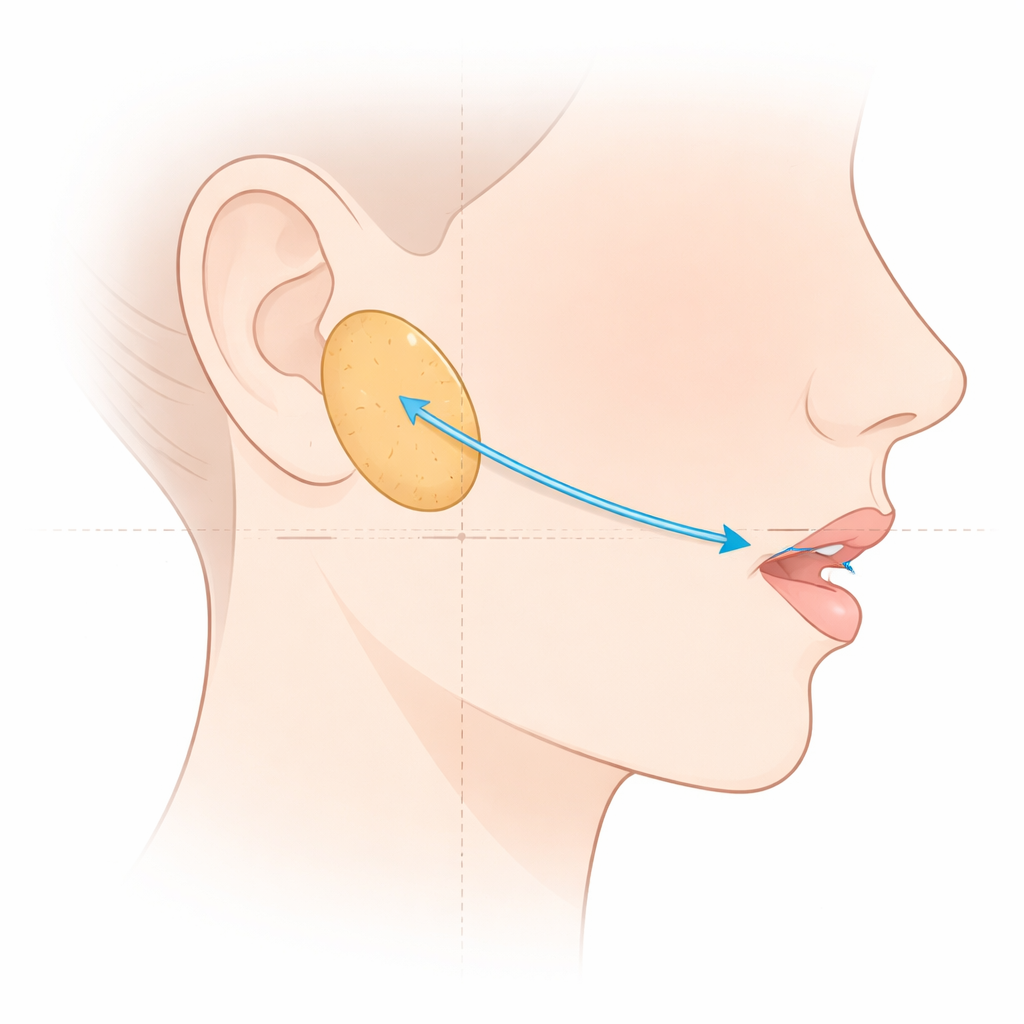
The parotid gland sits just in front of the ear and sends saliva into the mouth through the parotid duct, which travels across the chewing muscle in the cheek before turning inward through another muscle to reach the mouth cavity. Because this path is so close to the surface, doctors often use straight lines drawn between facial points—such as from the ear to the corner of the mouth—to guess where the duct lies under the skin. Past work showed these guesses were often good but mostly based on views from the side of the face. The authors of this study wanted a fuller three-dimensional picture, combining side and front views, and to see how well these surface rules could be checked with ultrasound in a living person.
Careful Mapping on Donated Bodies
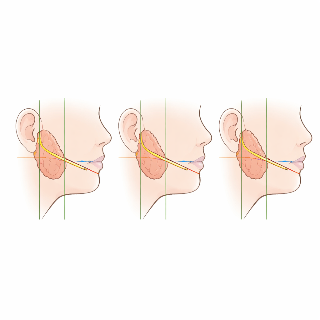
The researchers studied 32 sides of the face from 16 older adult donors. They exposed the parotid duct from where it leaves the gland to where it pierces the cheek muscle and enters the mouth. They then compared the duct’s position to three simple reference systems: a slanted line from the ear’s small flap (the tragus) to the corner of the mouth, horizontal levels across the upper lip groove between the nostrils and the lip (the philtrum), and vertical lines dropped from the inner and outer corners of the eye. At the point where the duct crossed the front edge of the chewing muscle, it lay just above the ear–mouth line in about five out of six specimens, right on the line in a few, and just below it in a small minority. Most of the “below the line” cases had a small extra parotid gland sitting in the cheek.
Front View Landmarks and Variations
From the front, the team looked at where the duct lined up as it crossed the cheek. In roughly two thirds of faces, it matched the mid-height of the upper lip groove, while in most of the remainder it sat slightly higher, and only rarely lower. Where the duct dove through the cheek muscle, it almost always did so a bit closer to the nose than a vertical line drawn straight down from the outer corner of the eye, and only exceptionally right under that corner. Overall, these patterns showed that, in spite of natural differences between people, the parotid duct keeps a fairly predictable course in relation to recognizable facial features, with deviations most likely when extra gland tissue is present.
Checking the Map with Sound Waves
To see whether this map would hold up in a living person, the authors used a high-resolution ultrasound machine on a healthy volunteer. Moving the probe along the same ear–mouth line, they were able to see the duct as a darker, tube-like structure just above that line at the front edge of the chewing muscle, mirroring what they had found in the cadavers. This ultrasound test was only done in one person, so it does not prove that the pattern is identical in everyone, but it does show that the duct’s relationship to simple surface lines can be visualized in real time, without any cutting.
What This Means for Everyday Care
For non-specialists, the key message is that the saliva duct in the cheek tends to run in a narrow, repeatable band that can be estimated using the ear, lips, and eye corners as guides. Knowing this helps surgeons, dentists, and cosmetic practitioners plan needle paths, thread lifts, or cuts that avoid the duct, and helps emergency doctors suspect and search for duct injuries when a facial wound crosses that zone. While the study was small and based mainly on older Korean donors, it offers a practical set of visual rules that, combined with ultrasound when needed, can make work on the midface safer and complications from unseen duct damage less likely.
Citation: Iwanaga, J., Tubbs, R.S., Kitagawa, N. et al. Facial landmark-based localization of the parotid duct: a cadaveric study with ultrasonographic feasibility assessment. Sci Rep 16, 11082 (2026). https://doi.org/10.1038/s41598-026-41701-1
Keywords: parotid duct, facial anatomy, ultrasound, cosmetic procedures, salivary gland