Clear Sky Science · en
Clinicopathological and imaging factors of surgical margin status and prognosis in breast-conserving therapy
Why this matters to women with breast cancer
For many women with breast cancer, keeping the breast through breast-conserving surgery (also called lumpectomy) is an attractive option. But this approach only works well when surgeons can remove the tumor completely while leaving healthy tissue behind. If cancer cells are found at the edge of the removed tissue, women may need another operation and may face a higher risk of the cancer returning in the breast or nearby lymph nodes. This study explores which medical scans and tumor features can best predict clean surgical edges and long-term control of the disease, helping doctors plan treatment that is both safer and less disruptive.
Looking closely at thousands of real-world cases
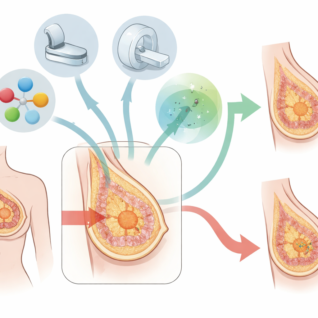
Researchers in Tianjin, China, reviewed the records of 2,775 women with invasive breast cancer who chose breast-conserving surgery between 2014 and 2024. Every patient had three imaging tests before surgery: ultrasound, mammography, and MRI. The team linked what they saw on these scans to detailed lab reports on the removed tumors and to follow-up information on whether the cancer later came back in the breast or nearby lymph nodes. In particular, they focused on whether cancer cells reached the cut edge of the tissue (a “positive margin”), whether women needed more than one breast-conserving operation, and which patients later developed local-regional recurrence.
Which factors signal trouble at the cutting edge
About one in five women in this study had positive margins after their first operation. Several features made this outcome more likely. Larger tumors and spread of cancer cells into tiny blood or lymph channels around the tumor were important warning signs. A heavy component of cancer growing within the milk ducts also strongly increased the chance of leftover disease. On imaging, ultrasound size was more predictive than mammography size, while MRI offered especially rich clues: a wider zone of abnormal enhancement around the tumor, denser breast tissue, stronger background enhancement in the surrounding breast, non-mass-like enhancing areas, and suspicious lymph nodes under the arm all pointed toward a higher risk of positive margins.
How tumor type and imaging shape surgical planning
Not all breast cancers behaved the same. Tumors driven by the HER2 molecule had the highest rate of positive margins, followed by certain hormone-sensitive types, while some triple-negative cancers had lower rates. Within each biological subtype, different imaging features mattered more. For example, in many hormone-sensitive cancers, the combination of a broader enhancing area on MRI and strong background enhancement around the tumor was especially telling. When the researchers compared the three imaging tests, ultrasound best matched the true size of the tumor seen in the lab, whereas MRI was the most accurate at judging which lymph nodes in the armpit were involved and at visualizing subtle spread of disease around the main mass. Among women who did have successful breast-conserving surgery, those who needed repeat operations were more likely to have HER2-positive tumors, extensive duct involvement, dense and strongly enhancing breast tissue on MRI, and suspicious lymph nodes.
Signals that predict cancer’s return close to home
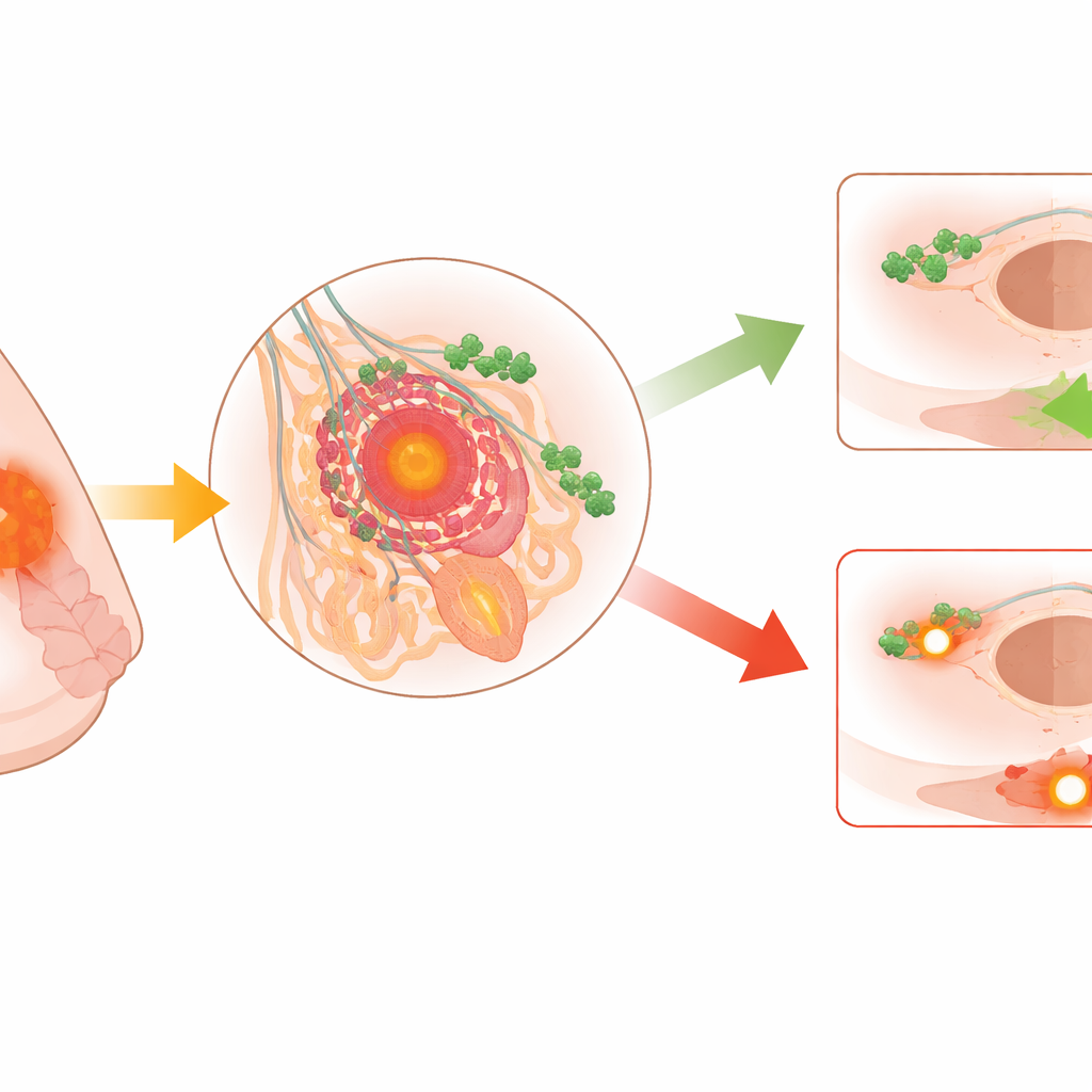
After a median of almost six years of follow-up, only 2.5% of women had the cancer return in the breast or nearby lymph nodes. Still, some groups carried much higher risk. Younger women, those with HER2-positive or triple-negative tumors, and those whose tumors invaded lymph or blood channels or grew extensively along the ducts faced more local-regional recurrences. MRI findings again proved informative: larger areas of abnormal enhancement and very strong background enhancement in the breast were tied to higher recurrence risk. Skipping radiation after surgery sharply increased the chance of the cancer coming back, underscoring the importance of this treatment even when the breast is preserved.
What this means for patients and doctors
This work suggests that combining modern imaging with detailed tumor typing can help doctors estimate in advance who is likely to need wider surgery, who may avoid repeat operations, and who needs especially close monitoring and strong follow-up treatment. Ultrasound gives a reliable sense of tumor size, while MRI helps reveal hidden spread in the breast and lymph nodes and highlights patients whose tissue patterns and tumor biology make clean margins harder to achieve. For women, this means that a tailored plan—built from scan findings and lab results—can improve the odds of keeping the breast, reduce the stress of unexpected re-operations, and lower the risk that cancer will return in the same area after treatment.
Citation: Liu, X., Liu, Y., Ma, T. et al. Clinicopathological and imaging factors of surgical margin status and prognosis in breast-conserving therapy. Sci Rep 16, 10450 (2026). https://doi.org/10.1038/s41598-026-41626-9
Keywords: breast-conserving surgery, breast MRI, surgical margins, HER2-positive breast cancer, local recurrence