Clear Sky Science · en
Principal component analysis of 3-dimensional facial soft-tissue morphology in three adult populations
Why the Shape of Our Faces Matters
When we look at a face, we instantly notice whether it seems balanced, expressive, or attractive, but it is hard to say exactly why. This study uses modern 3D cameras and advanced math to explore how the soft parts of the face—skin, fat, and muscles—differ among healthy adults from three ethnic groups. By turning detailed facial scans into simplified patterns, the authors hope to support more personalized orthodontic and surgical care that respects natural variation rather than forcing everyone to fit one narrow beauty ideal.
Looking at Faces in Three Dimensions
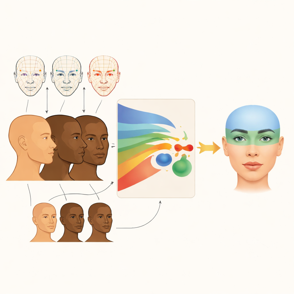
Instead of relying on flat photographs, the researchers worked with 210 three-dimensional facial scans of adults from Chinese, Hungarian, and Hispanic backgrounds, evenly split by sex. All participants had what orthodontists call “facially balanced” appearances: no major jaw problems, normal bite, healthy body weight, and no history of facial surgery or trauma. Using laser scanning and stereophotogrammetry systems, the team captured lifelike 3D models of each face under standardized lighting and head position so that subtle differences could be compared fairly.
Turning Faces into Measurable Patterns
To compare shapes, the researchers marked 57 key points on each face—on the forehead, around the eyes and nose, along the lips, and on the chin. These landmarks were checked for consistency and shown to be very reliable, usually within less than a millimeter. The faces were then digitally aligned so that they could be overlaid and compared. From there, the team used a technique called principal component analysis, which compresses many measurements into a few main patterns that explain most of the differences between people. In this case, just four such patterns captured more than three quarters of all the variation in soft-tissue facial shape.
Four Main Ways Faces Differ
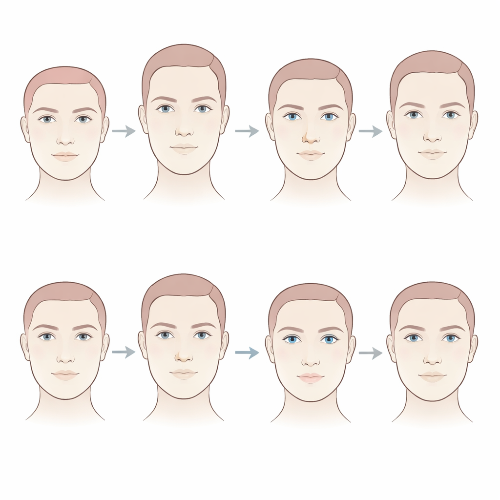
The first key pattern was overall height of the upper face—from the area near the eyebrows down toward the upper lip—which alone explained nearly half of all variation. Some people in the sample effectively had “taller” upper faces, while others had “shorter” ones. The second pattern described how far the nose projects compared with the position of the eyes: in some faces the nose tip sits farther forward relative to the eye area, in others it is closer to the plane of the eyes. The third pattern involved how far apart the eyes are from side to side and how high or low they sit relative to a point under the nose. The fourth pattern reflected how much the upper lip and the corners of the mouth project forward, capturing differences in lip fullness and mouth prominence.
Rethinking One-Size-Fits-All Beauty Rules
These four patterns were most pronounced in the upper half of the face, suggesting that healthy adults from different ethnic backgrounds resemble one another more in the lower face than in the forehead, eye, and upper lip regions. The findings challenge long-used “Neoclassical” facial rules, originally based on two-dimensional drawings of European faces, which are still sometimes used to judge harmony and guide treatment. The authors argue that applying those old standards to everyone can erase important aspects of individual and ethnic identity. Instead, 3D analysis of real, diverse faces offers a way to describe normal variation more accurately.
What This Means for Care and Aesthetics
For patients, the study’s message is that there is no single ideal face. Upper facial height, nose-to-eye balance, eye spacing, and lip prominence vary widely even among people considered attractive and well balanced. By using 3D imaging and mathematical tools to capture these patterns, orthodontists and surgeons can plan treatments that fit each person’s own facial structure and cultural background, rather than forcing their features toward outdated averages. The authors see this work as a first step toward more personalized, ethnically inclusive standards of facial harmony, to be refined in future studies using larger samples, long-term follow-up, and artificial intelligence.
Citation: Kau, C.H., Borbely, P., Zhurov, A. et al. Principal component analysis of 3-dimensional facial soft-tissue morphology in three adult populations. Sci Rep 16, 10316 (2026). https://doi.org/10.1038/s41598-026-41517-z
Keywords: 3D facial morphology, soft tissue face shape, ethnic facial variation, principal component analysis, personalized orthodontics