Clear Sky Science · en
Reliability assessment of the non-rigid registration for central incisors movement evaluation based on CBCT registration
Why this matters for your smile
Orthodontic treatment—braces and clear aligners—works by gently moving teeth into healthier, more attractive positions. To know whether treatment is really doing what it should, orthodontists must measure tiny tooth movements, often less than a millimeter. This study tests whether a newer, low-radiation X‑ray method can track those small changes as reliably as a more advanced 3D scan, without exposing patients to extra cost and radiation.
Two ways to look at the same teeth
Today’s orthodontists can choose between classic side‑view X‑rays of the head and jaws, called lateral cephalograms, and three‑dimensional cone‑beam CT scans that build a full 3D picture of the skull. The simple X‑ray is cheaper and uses less radiation, but it flattens a 3D skull into a 2D image and can slightly enlarge or distort structures. Cone‑beam CT avoids those distortions and offers more precise measurements, but at the price of higher radiation and cost. The central question of this research is whether the more common X‑ray can be upgraded with a smarter computer method so it performs almost as well as cone‑beam CT when tracking the movement of the upper and lower front teeth.
A smarter way to align X‑rays
To compare treatment before and after, images must first be “registered,” or lined up so that the skull, jaws and teeth are in the same reference position. The standard computer method for this task, known as iterative closest point, is good at lining up shapes but assumes they don’t change size and it is sensitive to noisy outlines. That is a problem for dental X‑rays, where the apparent size of structures can vary with machine settings or patient positioning, and where tooth and bone edges are not always crisp. Building on earlier work, the researchers tested a modified approach for the 2D X‑rays that allows slight uniform scaling to fix magnification differences and uses a statistical measure (maximum correntropy) to down‑weight misleading contour points. In practical terms, this algorithm lets the computer flexibly shrink, expand and clean up the X‑ray outlines so the before‑and‑after images match more faithfully.
Putting the new method to the test
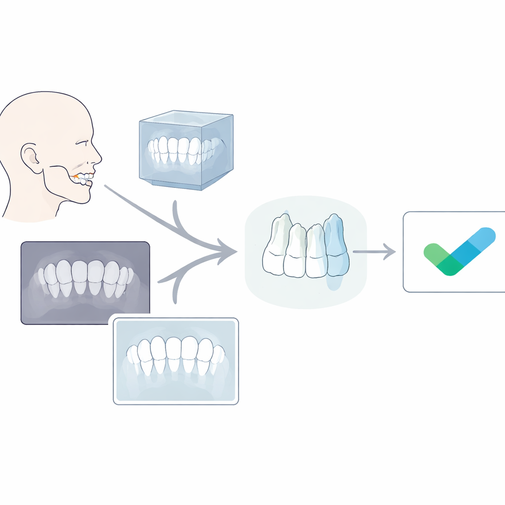
The team collected before‑and‑after records from 100 adult orthodontic patients, half of whom had four premolars removed as part of their treatment and half who did not. For each patient they had both side‑view X‑rays and cone‑beam CT scans taken before and after treatment. On the X‑rays, an orthodontist carefully traced key parts of the skull and jaws, including the upper and lower front teeth, and these outlines were matched using the new “non‑rigid” registration method. On the CT scans, the researchers reconstructed 3D skull models and used well‑defined anatomical points in the upper face and lower jaw to align the before‑and‑after scans. In both kinds of images, they then measured how far the tips of the upper and lower central incisors had moved forward or backward.
How close were the measurements?
When the researchers compared tooth movement measured from the improved X‑ray method with the same movement measured from the 3D scans, the differences were consistently small—typically between about half and three‑quarters of a millimeter. Statistical tests showed no meaningful differences between the two methods for either the upper or lower front teeth, regardless of whether patients had premolars removed. In other words, the upgraded 2D technique and the 3D cone‑beam CT agreed closely enough that, from a clinical standpoint, they told the same story about how far the incisors had moved.
What this means for patients and clinicians
The study concludes that a carefully designed computer algorithm can make routine side‑view X‑rays reliable for tracking front‑tooth movement, rivaling the accuracy of 3D cone‑beam CT for this purpose. That matters because X‑rays are cheaper, faster and expose patients to less radiation, yet with this method they still provide precise feedback on how well treatment is working. While the approach currently focuses on adults and mainly captures movement in up‑and‑down and front‑to‑back directions, it already offers orthodontists a practical, safer tool to monitor tooth movement without routinely resorting to higher‑dose 3D imaging.
Citation: Wu, Zx., Shi, Zy., Bu, Wq. et al. Reliability assessment of the non-rigid registration for central incisors movement evaluation based on CBCT registration. Sci Rep 16, 12957 (2026). https://doi.org/10.1038/s41598-026-41254-3
Keywords: orthodontic imaging, tooth movement, cephalometric X-ray, cone-beam CT, image registration