Clear Sky Science · en
Integrated multi-omics analysis combined with clinical validation reveals that HLA-DRB5 and ODAPH are causal risk genes for keratoconus
Why this eye disease matters
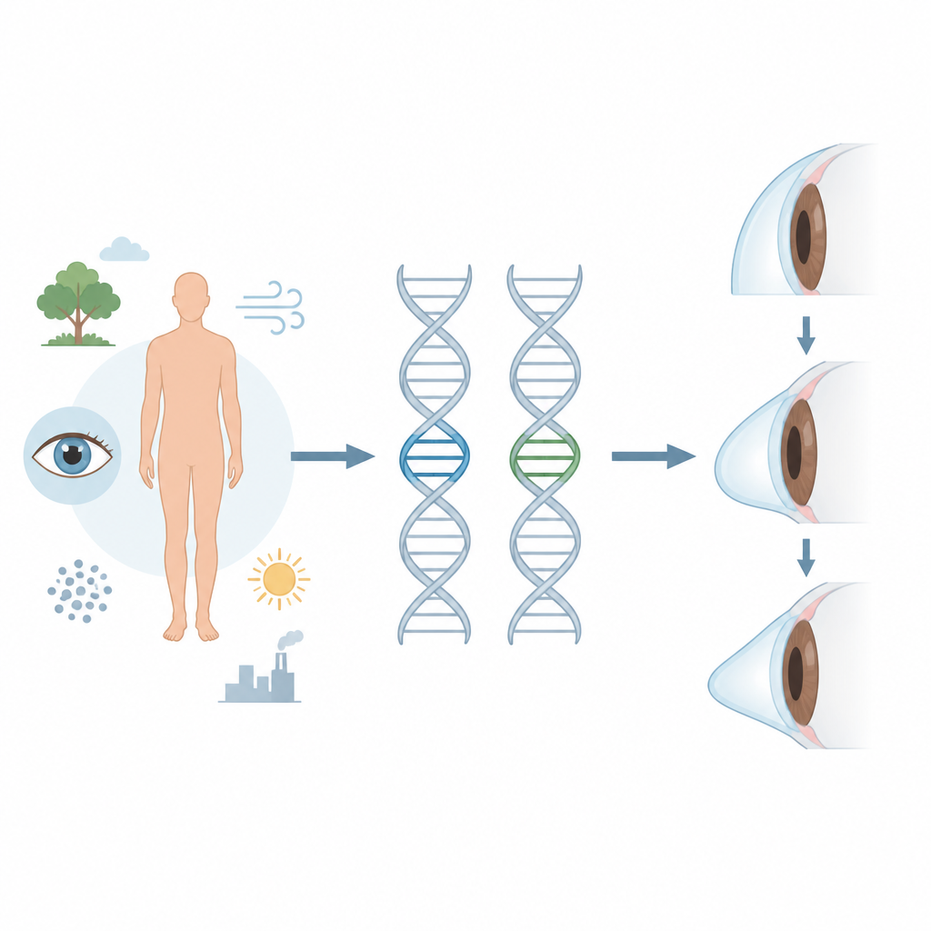
Keratoconus is a disorder in which the clear front window of the eye gradually thins and bulges into a cone, often starting in the teenage years. It can blur vision, cause sensitivity to light, and sometimes lead to corneal transplants. Current treatments mainly manage symptoms rather than the root cause. This study looks deep into human DNA and tissue samples to pinpoint which genes help drive keratoconus, opening the door to earlier detection and more targeted care.
Looking at the eye from the gene level
To understand what goes wrong in keratoconus, the researchers combined several layers of biological data, a strategy often called multi omics. They analyzed large public datasets of gene activity in corneal tissue from people with and without keratoconus, then matched these with wide scale genetic studies that link DNA differences to disease risk. Instead of simply asking which genes look different, they asked which differences are likely to play a causal role in the disease, not just accompany it.
Finding suspect genes in a crowded field
From two independent cornea datasets, the team identified 2,884 genes that were more active in keratoconus than in healthy corneas. Many of these genes are involved in how cells stick to their surroundings and how the immune system reacts, as well as in well known inflammatory signaling routes. Using statistical approaches that treat genetic variants as natural experiments, they then tested whether changes in gene activity are likely to influence the chance of developing keratoconus. This filtering step narrowed the list to a smaller group of genes with strong signs of causal involvement.
Two stand out genes and how they may harm the cornea
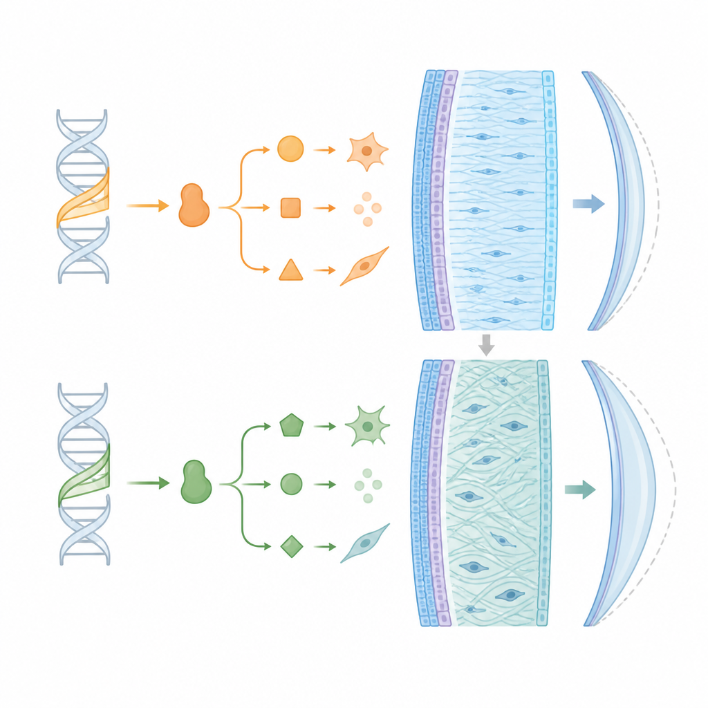
Among the many candidates, two genes stood out: HLA DRB5 and ODAPH. Variants that increase their activity were strongly tied to a higher risk of keratoconus. HLA DRB5 is best known for its role in directing immune responses. Overactivity of this gene may disturb the eye's normally quiet immune environment, feeding ongoing low level inflammation and activating enzymes that break down the corneal structure. ODAPH, first studied in tooth enamel formation, appears to help control how tissue building blocks stick together and harden. In the cornea, abnormal ODAPH activity may weaken the collagen framework that keeps the cornea smooth and dome shaped, making it more prone to thinning and bulging.
Testing real tissues and hinting at new treatments
To move beyond computer analyses, the researchers examined corneal tissue and blood from five keratoconus patients and five control donors. They found that HLA DRB5 and ODAPH, along with a tissue breaking enzyme called MMP 9, were significantly more active in both the patients' corneas and their blood. This agreement between genetic statistics and real world samples strengthens the case that these genes are not just bystanders. The team also searched drug databases and highlighted an experimental antibody, Meplazumab, which targets the pathway linked to HLA DRB5 and has already been tested in other conditions, although it still faces challenges in reaching corneal tissue effectively.
What this means for people with keratoconus
This work suggests that HLA DRB5 and ODAPH are key risk genes that help drive keratoconus, likely by disturbing immune balance and weakening the corneal scaffold. For patients, that does not translate into an immediate new treatment, but it offers a clearer map of the disease. These genes could form the basis of future blood or tissue tests to spot high risk individuals earlier, refine risk scores, or monitor disease activity. Over time, medicines that calm the harmful immune signals or protect the corneal structure by acting on these gene driven pathways may complement existing procedures, bringing care closer to the root causes of keratoconus.
Citation: Zhao, F., Zhao, B., Cao, R. et al. Integrated multi-omics analysis combined with clinical validation reveals that HLA-DRB5 and ODAPH are causal risk genes for keratoconus. Sci Rep 16, 15185 (2026). https://doi.org/10.1038/s41598-026-41037-w
Keywords: keratoconus, corneal genetics, multi-omics, immune pathways, collagen thinning