Clear Sky Science · en
Propofol and dexmedetomidine sedation share the similar functional activity but distinct functional synchronization
Why this matters for everyday anesthesia
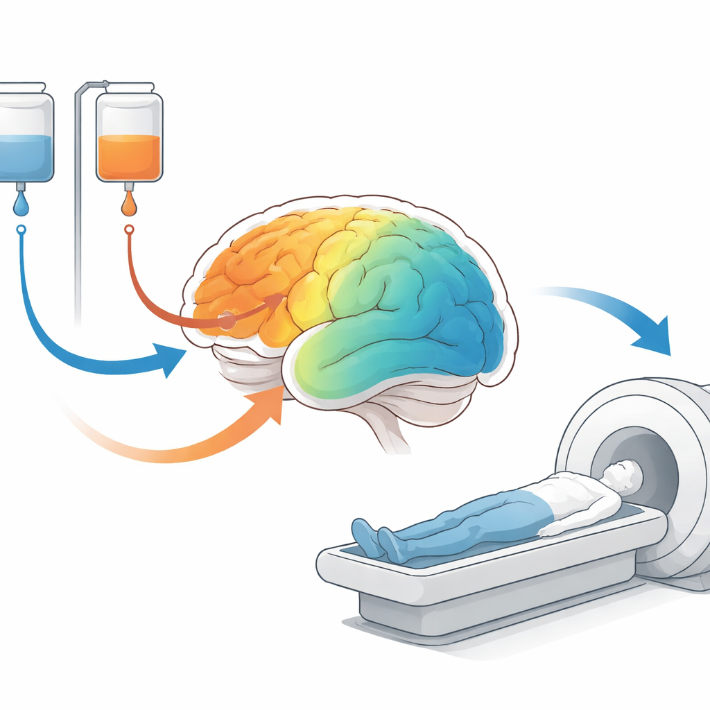
Each year, millions of people receive anesthesia for surgery or medical scans, yet we still do not fully understand how these drugs switch consciousness off and on. This study looks inside the resting human brain with MRI while two common sedative drugs, propofol and dexmedetomidine, are given to healthy volunteers. By comparing how brain activity and communication between regions change, the researchers show that the two drugs quiet and reshape the brain in similar ways locally, but disturb its internal communication in different ways. These insights may ultimately help make anesthesia safer and more precisely tailored to individual patients.
Two drugs, one question
Propofol and dexmedetomidine are widely used but act on very different chemical systems: propofol boosts the brain’s main inhibitory signal, while dexmedetomidine taps into natural sleep pathways. To compare their effects fairly, the researchers recruited 42 healthy adults and divided them into two groups. One group received propofol and was scanned in four states: awake, mildly sedated, deeply sedated, and recovered. The other group received dexmedetomidine and was scanned while awake, mildly sedated, and recovered. During each state, the team used resting-state functional MRI, which measures slow, spontaneous fluctuations in brain blood flow while people lie still in the scanner.
How the brain’s local activity shifts
The authors first asked how strongly different brain areas were “idling” during rest. They used two standard measures: one that tracks the strength of slow signal fluctuations and another that tracks how synchronized neighboring spots are. Despite their chemical differences, both drugs produced a strikingly similar pattern as sedation deepened. Activity in the frontal lobes, which support planning, attention, and conscious awareness, consistently dropped. In contrast, regions along the side and top of the brain, such as the superior temporal gyrus and a band of tissue that helps control movement and bodily sensations, showed increased local activity and synchronization. Some visual regions at the back of the brain briefly became more active at light doses, especially with dexmedetomidine, but this effect faded at deeper levels of propofol sedation.
How brain networks fall out of step
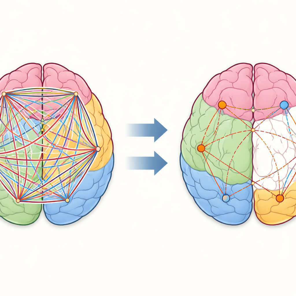
Next, the team examined how well distant brain regions rose and fell together over time, a measure known as functional connectivity. Here, propofol and dexmedetomidine diverged. Under propofol, the first connections to weaken linked networks involved in moving the body and shifting attention. As the drug level increased, connections within and between several major networks weakened, including those that support goal-directed control and the so-called “default” network that is active during daydreaming and self-reflection. Yet not all links were lost: a small subset of connections within and between these higher networks actually grew stronger, hinting that the sedated brain may reorganize rather than simply shut down.
A different kind of disconnection
Dexmedetomidine told another story. At mild sedation, connectivity across almost the entire brain dropped in a more uniform way, without the pattern of selective increases seen with propofol. This broad dampening fits with prior work suggesting that dexmedetomidine makes brain networks less efficient overall, more like a deep but natural sleep. For both drugs, the brain’s communication patterns in the recovery scans looked statistically similar to the awake state, even though some measures of local activity still shifted between deep sedation and recovery. This suggests that different aspects of brain function may rebound on different timescales as people wake up.
What this means for understanding unconsciousness
To a non-specialist, the main message is that two very different anesthetic drugs nudge the resting brain into a similar pattern of local quieting in the frontal lobes and heightened activity in certain sensory and movement regions. Where they differ is in how they disrupt the brain’s long-distance conversations: propofol selectively weakens and reshapes communication among key control and “default” networks, while dexmedetomidine produces a more widespread, even dampening of connections. These complementary patterns support the idea that losing consciousness is not caused by a single “off switch,” but by changes in both how strongly different areas fire and how well they stay in sync. Mapping these changes more precisely may help anesthesiologists better monitor depth of sedation and could shed light on other conditions where awareness is altered, from sleep to coma.
Citation: Minyu, J., Jiayi, Z., Guiyu, L. et al. Propofol and dexmedetomidine sedation share the similar functional activity but distinct functional synchronization. Sci Rep 16, 11768 (2026). https://doi.org/10.1038/s41598-026-40974-w
Keywords: anesthesia, propofol, dexmedetomidine, brain connectivity, resting-state fMRI