Clear Sky Science · en
A lightweight transformer-based hybrid encoder-decoder model for chest X-ray medical report generation
Why faster chest scans matter
Chest X-ray scans are one of the most common ways doctors look for lung and heart problems, but turning each image into a clear written report takes time and concentration. In busy hospitals or remote clinics, specialists are often overloaded, which can delay treatment and increase the chance of mistakes. This study presents a computer system called FAST-MRG that looks at a chest X-ray and automatically drafts a full paragraph-style report, similar to what a radiologist would write. The goal is not to replace doctors, but to give them a fast, reliable first draft that can speed up care and bring expert-style reporting to places with few specialists.
From picture to paragraph
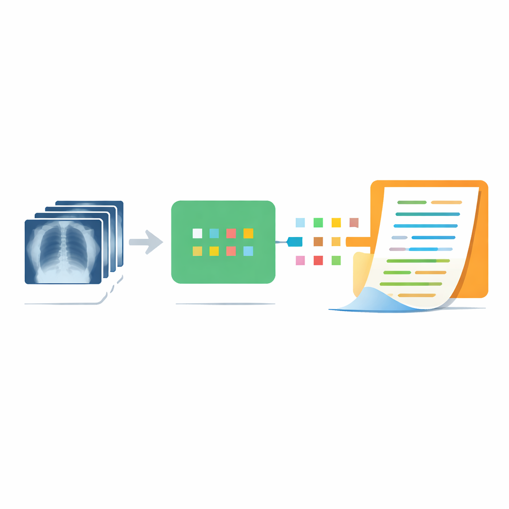
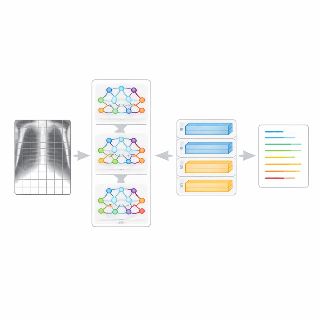
The core idea behind FAST-MRG is to connect two powerful kinds of artificial intelligence: one that is good at understanding images and another that is good at writing text. On the image side, the system breaks each chest X-ray into many small patches and analyzes how they relate to one another, rather than scanning the picture line by line. On the text side, it uses a language model that has learned how words flow together in natural paragraphs. By linking these parts, FAST-MRG takes in a single chest X-ray and outputs a multi-sentence description of what the image shows, much like the “findings” and “impression” sections that doctors type into medical records.
Learning from real hospital reports
To train and test the system, the researchers used the Indiana University Chest X-Ray Collection, a widely used public dataset. It contains 6,469 chest X-ray images paired with real reports written by radiologists. These reports vary in length, word choice, and style, reflecting the way different doctors actually write under real-world pressures. Because the wording is not standardized, teaching a computer to match these paragraphs is far harder than teaching it to choose a single disease label. The team carefully prepared the data, cleaning up obvious inconsistencies in spelling and punctuation while preserving genuine medical wording so the system would learn to operate in realistic hospital conditions.
A nimble brain for images and words
FAST-MRG is designed to be lightweight, meaning it can run relatively quickly and with modest computing power. For the image side, it uses a modern “transformer” model that has been taught to imitate a stronger teacher network, a process known as distillation. This allows the system to learn rich visual patterns from a limited medical dataset without needing huge amounts of training time. For the text side, it uses a transformer-based language model that builds the report one word at a time, always taking into account what has already been written so that the paragraph stays fluent and medically sensible. Together, these choices let the system balance accuracy with speed, which is crucial if it is to be useful in real clinics.
How well the system performs
The researchers compared FAST-MRG with earlier methods that also try to turn chest X-rays into text. Using standard measures of how closely computer-generated text matches human-written reports, FAST-MRG produced better multi-word phrases and more natural sentences than most competing systems. It especially shined on tests that reward getting longer fragments of language correct, which suggests it is good at capturing full ideas rather than just isolated terms. At the same time, the model trained significantly faster than many heavier designs that rely on bulkier image networks. Detailed charts showed that the system’s performance was steady across hundreds of test cases, with few extremely bad outputs, an important property for any tool that might one day support clinical work.
What this means for patient care
For a non-specialist, the key message is that computers are getting better at translating complex medical images into coherent, paragraph-style language, and FAST-MRG is a promising step in that direction. The system can draft meaningful reports in seconds, helping doctors focus on judgment rather than routine description, and offering a safety net in crowded or under-staffed settings. The authors stress that such tools should be used as decision support, with human experts always reviewing the output, especially because rare conditions and subtle findings remain challenging. Even so, the study shows that carefully designed, efficient AI systems can bring high-quality reporting closer to every patient, and the same ideas could eventually extend to scans of the brain, abdomen, and other parts of the body.
Citation: Ucan, M., Kaya, B., Kaya, M. et al. A lightweight transformer-based hybrid encoder-decoder model for chest X-ray medical report generation. Sci Rep 16, 8645 (2026). https://doi.org/10.1038/s41598-026-40710-4
Keywords: chest X-ray, medical report generation, transformer models, clinical decision support, radiology AI