Clear Sky Science · en
Single cell multiomic landscape reveals gene programs driving lipid droplet heterogeneity in hepatic steatosis
Why this matters for liver health
Many people who drink heavily develop a “fatty liver” long before they notice symptoms. Sometimes this early change quietly progresses to cirrhosis and liver failure, but doctors still do not fully understand why. This study combines advanced single-cell and imaging technologies to show, in unprecedented detail, where fat builds up inside the liver, which cell programs drive it, and how specific genes and regulatory switches may tip the balance toward disease—or protection.
Where fat collects inside the liver
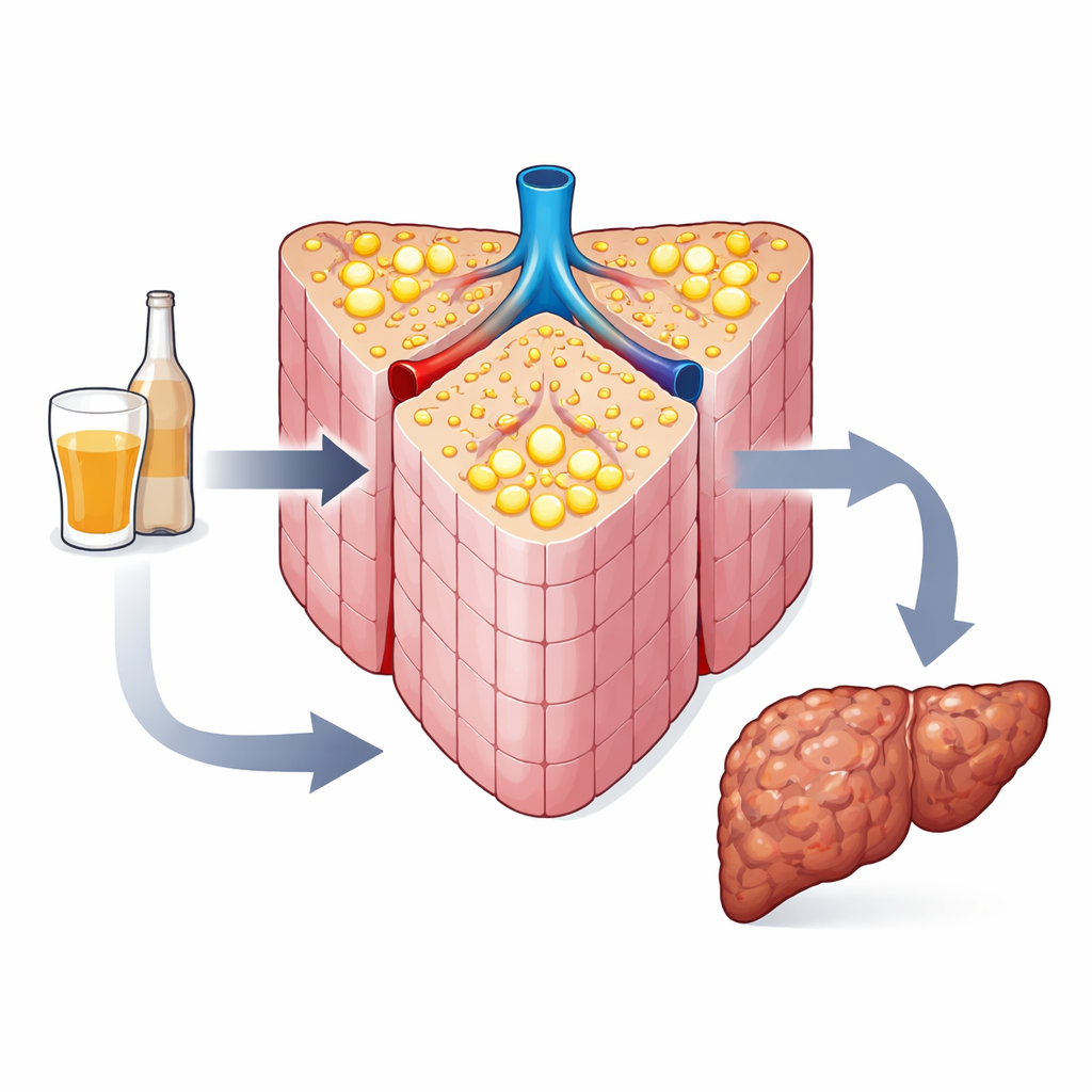
The liver is organized into tiny repeating units called lobules, each stretching from incoming blood vessels at the portal region to an outgoing central vein. The authors analyzed human liver biopsies from people with alcohol-associated liver disease (ALD) and compared them to biopsies from patients with metabolic fatty liver disease. Using a machine-learning algorithm trained by expert pathologists, they mapped and measured hundreds of thousands of fat droplets within these lobules. In ALD, fat droplets were not spread evenly: they were more numerous and larger near the portal regions, whereas in metabolic disease they mainly clustered near the central veins. This revealed that alcohol creates a distinct, zone-specific pattern of fat storage that standard microscopy had missed.
Confirming the pattern in experimental models
To probe mechanisms, the team turned to mice fed alcohol for eight weeks, a model of early human ALD. Classic staining methods and fluorescent labeling of fat confirmed that, as in patients, mouse livers developed many more and larger droplets in the periportal areas than near central veins. By co-labeling known landmark proteins, the researchers could reliably orient each image along the portal–central axis and quantify droplet number and size in each zone. These experiments showed that periportal dominance of fat accumulation is a robust feature of alcohol-induced liver injury across species.
Zooming in on cell programs and DNA access
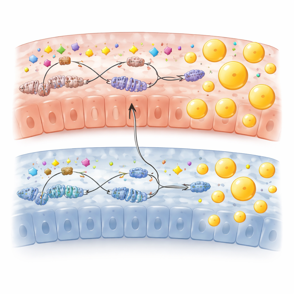
The investigators then asked what liver cells in different zones were actually doing. Using single-cell RNA sequencing, they profiled thousands of individual hepatocytes from alcohol-fed and control mice and computationally reconstructed their positions along the portal–central axis. More than half of all liver genes showed non-random spatial patterns, and alcohol created new zone-specific programs. In periportal hepatocytes, genes involved in building and handling fats and cholesterol, including Fasn, Scd1 and a lesser-known gene called Hsd17b13, were strongly upregulated. In contrast, pericentral cells remained focused on drug breakdown. Parallel single-cell chromatin profiling (scATAC-seq) showed that the “open” regions of DNA and the activity of transcription factors—the proteins that turn genes on and off—also differed by zone and were reshaped by alcohol exposure.
A key gene and its molecular switches
Among the lipid-related genes, HSD17B13 stood out. Genetic studies in humans had previously linked loss-of-function variants in this gene to lower risk of alcoholic cirrhosis, but its in vivo regulation was unclear. Here, Hsd17b13 was one of the most upregulated genes in alcohol-fed mice and was almost exclusively expressed in periportal hepatocytes. High-resolution RNA imaging confirmed this spatial restriction, and protein studies showed that HSD17B13 sits directly on lipid droplets, especially in alcohol-exposed livers. Using chromatin accessibility data and 3D DNA-contact assays, the authors identified an alcohol-responsive enhancer that physically loops to the Hsd17b13 promoter. They further showed that two transcription factors, HNF4α (enriched periportally) and PPARα (more active pericentrally), bind both promoter and enhancer, forming a spatial “tug-of-war” that helps determine how much HSD17B13, and thus how much fat storage, occurs in each zone.
What this means for patients and therapies
Altogether, the study portrays early alcohol-associated fatty liver not as a uniform coating of fat, but as a finely patterned, periportal-centered process driven by zone-specific gene programs and chromatin states. Periportal hepatocytes, pushed by HNF4α and related factors, ramp up fat production and store it in large droplets enriched in HSD17B13, while pericentral cells emphasize fat burning and detoxification. This spatial imbalance may set the stage for later inflammation, scarring and cirrhosis. By pinpointing molecules like HSD17B13 and its upstream switches as key players in specific liver zones, the work suggests new, more precise targets for preventing or slowing alcohol-related liver damage before irreversible scarring takes hold.
Citation: Sehrawat, T.S., Cooper, S.A., Navarro-Corcuera, A. et al. Single cell multiomic landscape reveals gene programs driving lipid droplet heterogeneity in hepatic steatosis. Sci Rep 16, 10219 (2026). https://doi.org/10.1038/s41598-026-39913-6
Keywords: alcoholic fatty liver, hepatic steatosis, single cell multiomics, lipid droplets, HSD17B13