Clear Sky Science · en
White matter microstructure differences in obstructive sleep apnea severity groups assessed by diffusion tensor metrics and biophysical modeling
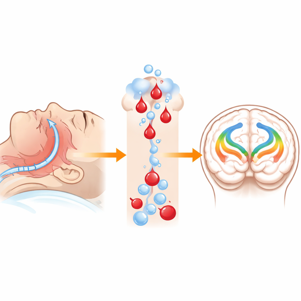
Why Trouble Breathing at Night Matters for the Brain
Many older adults think of loud snoring and pauses in breathing during sleep as just an annoyance. This study shows that obstructive sleep apnea, a condition marked by repeated airway blockages at night, may quietly reshape key wiring inside the brain even before obvious memory problems appear. By using advanced brain scans, the researchers looked beneath the surface to see how sleep apnea of different severities affects the brain’s white matter—the bundles of fibers that let different regions talk to each other.
Looking Beneath the Brain’s Wiring
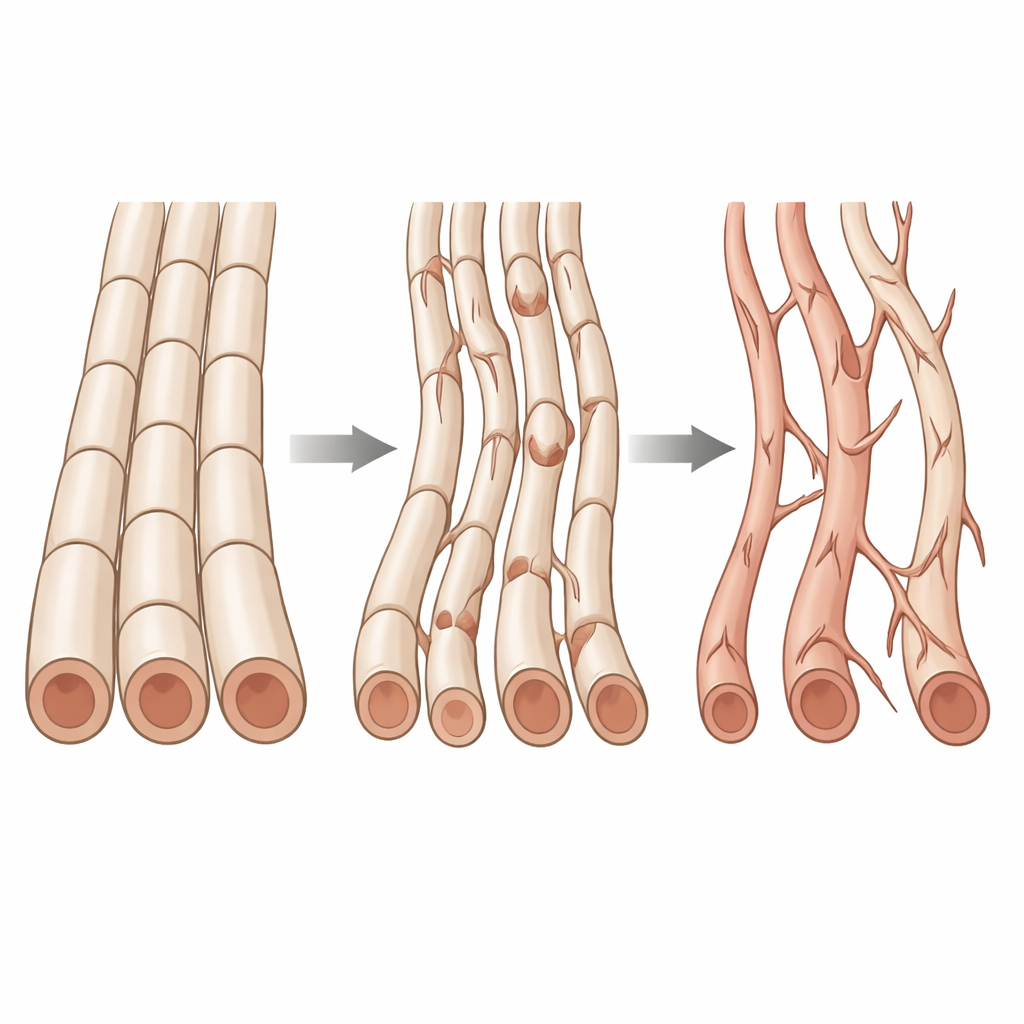
White matter is made of long nerve fibers that carry signals between brain regions, much like cables linking cities. The team studied 150 adults in their 60s and early 70s who were carefully screened to ensure they had normal thinking and no dementia. Each participant spent a night in a sleep laboratory, where their breathing, oxygen levels, and brain waves were monitored to measure how often their airway collapsed during sleep. They also underwent high‑resolution MRI scans designed to detect very subtle changes in the structure of white matter.
New Ways to Read Water’s Motion
Water molecules move differently in healthy brain tissue than in tissue that has been damaged or changed by disease. The researchers used several diffusion MRI methods that track this microscopic motion. Traditional measures, like diffusion tensor imaging, capture how freely water flows and in which directions. More advanced approaches, including diffusion kurtosis imaging and a biophysical "Standard Model," help tease apart whether changes are likely due to loss of insulation around fibers, damage to the fibers themselves, or shifts in the space between them. Together, these tools act like a set of complementary lenses on the same wiring.
Sleep Apnea Severity and Brain Hotspots
When the scientists compared people with little or no sleep apnea to those with mild, moderate, or severe disease, they found the clearest differences in three major white matter pathways. The front portion of the corpus callosum—which links the left and right frontal lobes—the cingulum, which connects regions important for attention and memory, and the external capsule, involved in broader communication networks, all showed changes as apnea worsened. In these tracts, people with more severe apnea tended to have diffusion patterns consistent with reduced fiber integrity and altered tissue organization.
Signs of Fiber Loss and Sheath Damage
The more often participants stopped breathing during sleep, the more their scan signals suggested thinning of the fatty coating that insulates nerve fibers (myelin) and a loss of the fibers themselves. Measures linked to how tightly water is constrained along healthy, bundled fibers decreased, while measures tied to water spreading outward increased. A particularly informative metric that reflects the fraction of water inside axons—the core of the nerve fibers—was lower in people with more severe apnea. These patterns fit with a picture of both damage to the insulating sheath and loss or distortion of the fibers, likely driven by repeated drops in oxygen and related inflammation. Some of these effects differed between men and women, hinting that sex may shape vulnerability or disease course.
What This Means for Everyday Thinking
Even though everyone in the study still scored as cognitively normal, the affected white matter regions are known to support memory and executive skills such as planning and decision‑making. The findings suggest that obstructive sleep apnea may quietly erode brain wiring in these areas long before noticeable cognitive decline sets in. In plain terms, nightly breathing problems are not just a sleep issue; they may be a slow‑acting stressor on the brain’s communication cables. The authors argue that following people over time, and tracking how apnea treatment changes these brain signals, will be crucial to understanding whether early detection and intervention can help preserve brain health and delay conditions like dementia.
Citation: Figueredo, L.F., Chen, J., Gaggi, N.L. et al. White matter microstructure differences in obstructive sleep apnea severity groups assessed by diffusion tensor metrics and biophysical modeling. Sci Rep 16, 11963 (2026). https://doi.org/10.1038/s41598-026-39162-7
Keywords: obstructive sleep apnea, white matter, brain MRI, cognitive aging, sleep and memory