Clear Sky Science · en
Distinct functional profiles of oral neutrophils in molar incisor pattern periodontitis, generalized periodontitis and periodontal health
Why the body’s first responders matter for gum health
Gum disease is not only about bacteria on our teeth; it is also about how our own immune system reacts. This study looks at neutrophils—white blood cells that act as first responders—to see how they behave in people with different forms of periodontitis compared with those with healthy gums. Understanding these differences may help explain why some people lose bone around their teeth quickly, while others remain stable, and could open the door to more precise ways to diagnose and treat gum disease.
Different types of gum disease, different stories
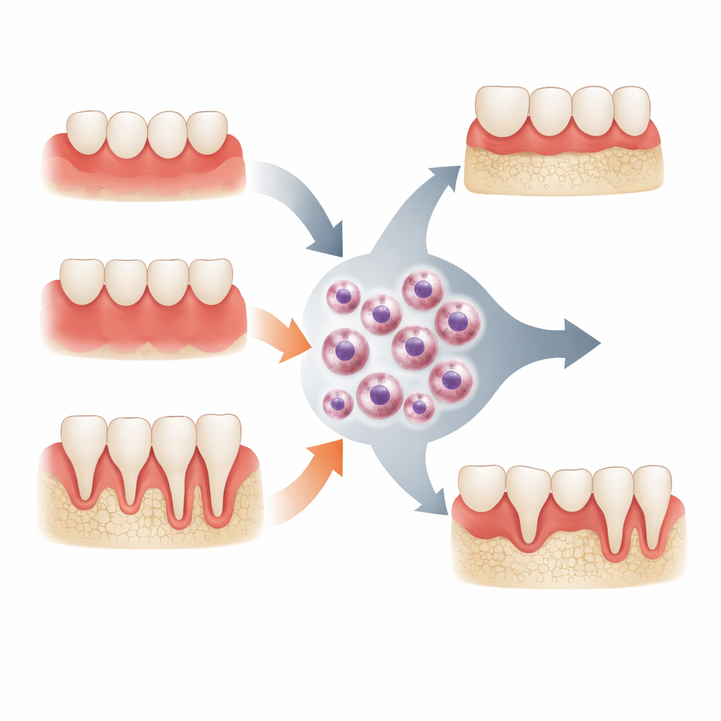
Periodontitis is a chronic infection of the tissues that hold teeth in place, leading to loss of the supporting bone. The authors focus on two forms: generalized periodontitis, which affects many teeth over time, and a more localized, fast-moving form that targets molars and incisors. Both are linked to an imbalanced community of microbes living under the gumline, but their microbial make-up differs. In particular, the more aggressive molar–incisor pattern is strongly tied to a highly toxic strain of a bacterium called Aggregatibacter actinomycetemcomitans. The study asks whether these distinct microbial communities drive equally distinct behaviors in oral neutrophils, the neutrophils that have already traveled through gum tissue and into saliva.
Sampling the mouth’s first line of defense
The researchers collected saliva rinses from three groups of volunteers: ten with healthy gums, ten with generalized periodontitis, and ten with molar–incisor pattern disease. From these rinses, they isolated oral neutrophils and tested how many cells were present, how robustly they produced reactive oxygen species (ROS, chemically reactive molecules used to kill microbes), whether they tended to die by necrosis, and which signaling molecules (cytokines) they released. The cells were challenged in the lab with the aggressive JP2 clone of A. actinomycetemcomitans, or with a chemical stimulus, and were compared with untreated cells. In a separate set of experiments, oral neutrophils from healthy donors were exposed to simplified biofilms that mimicked the microbial communities of health, generalized disease, or molar–incisor disease.
Weakened firepower and skewed signals
People with both types of periodontitis had more oral neutrophils in their saliva than healthy participants, reflecting the heavy traffic of immune cells into inflamed gum pockets. Yet, despite this higher number, their cells showed weaker oxidative “burst” activity: across conditions, neutrophils from healthy mouths produced more ROS than those from either disease group. When exposed to the JP2 bacterial strain, neutrophils from periodontitis patients, especially from the molar–incisor group, struggled to mount a strong ROS response and showed patterns consistent with increased necrosis. The team also found that neutrophils from the aggressive molar–incisor form tended to release more pro-inflammatory and bone-resorbing signals, such as TNFα and RANKL, and less of the protective mediator OPG, leading to a lower OPG/RANKL balance that favors bone breakdown.
How dental plaque communities shape neutrophil behavior
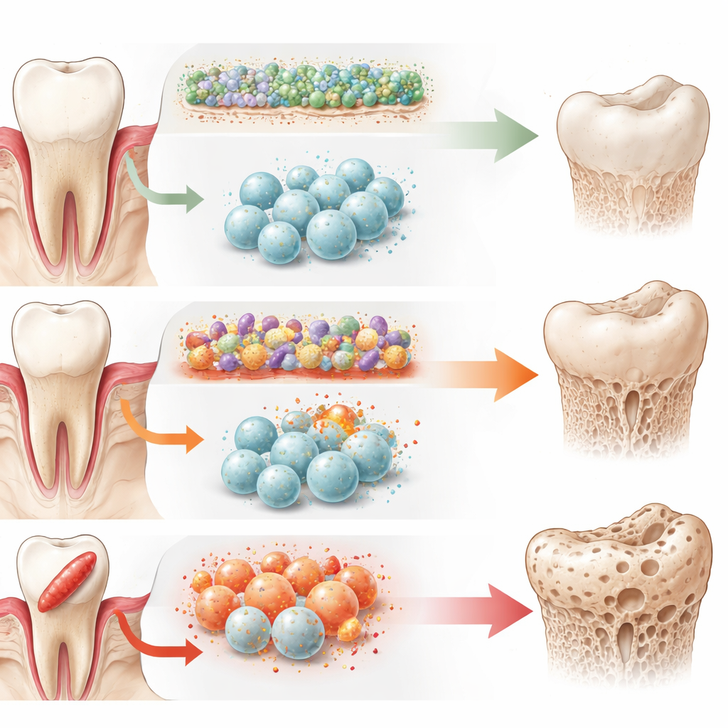
To tease apart the influence of microbes themselves, the researchers exposed healthy volunteers’ oral neutrophils to laboratory-grown biofilms modeled on either healthy or diseased gums. Under biofilms representing diseased states, neutrophils surprisingly produced less ROS but secreted higher levels of inflammatory and bone-affecting mediators, including TNFα, RANKL, and OPG, than they did under the healthy biofilm. This suggests that dysbiotic plaque may “reprogram” neutrophils to shift from a mainly microbe-killing role toward one that powerfully shapes the surrounding tissue environment, including bone remodeling. Importantly, these biofilm-driven changes did not fully match the profiles seen in patients, implying that long-term host factors and chronic exposure in the mouth further tune neutrophil behavior.
What this means for keeping teeth and bone
Overall, the study shows that oral neutrophils carry distinct functional fingerprints that mirror the health of the gums and the type of gum disease present. In periodontitis, these cells are more numerous but less effective at mounting oxidative attacks against key bacteria, while at the same time sending stronger signals that encourage bone loss—particularly in the fast-moving molar–incisor form. This dual shift, from efficient defenders to drivers of inflammation and bone resorption, helps explain why some patients experience rapid, localized destruction around certain teeth. In the future, measuring oral neutrophil patterns in saliva could aid early diagnosis, refine risk assessment, and guide therapies aimed not only at removing plaque but also at calming or redirecting the body’s own immune response.
Citation: Khoury, J., Haloun, B., Musai, N. et al. Distinct functional profiles of oral neutrophils in molar incisor pattern periodontitis, generalized periodontitis and periodontal health. Sci Rep 16, 12641 (2026). https://doi.org/10.1038/s41598-026-39112-3
Keywords: oral neutrophils, periodontitis, gum inflammation, oral biofilm, bone loss