Clear Sky Science · en
Do mandibular functional units exhibit inherent asymmetry in adults with a normal degree of chin deviation? A cone-beam computed tomography study
Why our jaws are never perfectly even
Look closely in the mirror and you may notice that your face is not perfectly the same on both sides—and that is normal. Dentists and orthodontists, however, need to know how much unevenness inside the jaw is "built in" and how much signals a real problem that might affect chewing, comfort, or appearance. This study used three‑dimensional scans to explore how the lower jaw (the mandible) differs from side to side in adults whose chins look straight, revealing hidden patterns of imbalance and compensation inside the bone.
Hidden unevenness behind a straight smile
Facial beauty is often linked with balance, and obvious jaw unevenness can change how the chin lines up with the center of the face, sometimes leading to bite problems and discomfort. But most people seen in orthodontic clinics have only tiny chin shifts—too small for friends or family to notice. The question the researchers asked was: even when the chin looks centered, do the left and right halves of the jaw still differ in size and shape? And does that hidden unevenness depend on how the upper and lower jaws fit together from front to back, a feature specialists classify into three broad skeletal types (Classes I, II, and III)?
High‑detail 3D views of the jaw
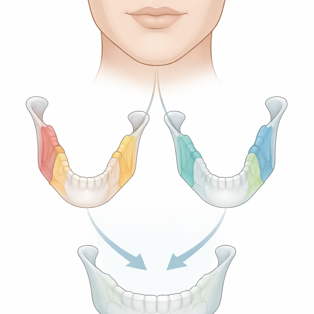
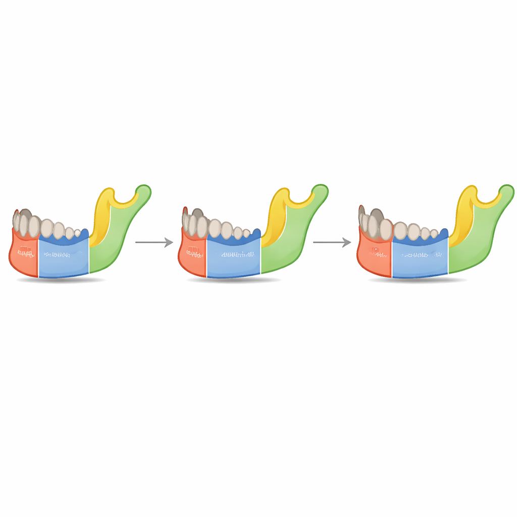
To answer these questions, the team analyzed cone‑beam computed tomography (CBCT) scans from 90 young adults who all had nearly straight chins and a similar vertical facial pattern. Using specialized software, they converted each scan into a three‑dimensional model of the mandible. They then digitally sliced each jaw into two halves and further into seven functional pieces, or "units": the condyle, coronoid, ramus, angle, body, alveolar region that holds the teeth, and the chin. For each unit on the left and right sides, they measured both length and volume, and calculated an asymmetry index that expresses how different the two sides are as a percentage.
Uneven on the inside, balanced on the outside
The results showed that none of the jaw units were perfectly matched. Across all participants and all skeletal classes, the average asymmetry index for every length and volume was clearly greater than zero, meaning some degree of built‑in unevenness is the rule, not the exception. Differences in volume tended to be larger than differences in simple linear distances, highlighting that three‑dimensional measurements capture subtle imbalances better than one‑dimensional ones. Among all units, the coronoid region and the angled corner of the jaw showed the greatest unevenness, while the overall half‑jaw volume and the full length along the lower border of the jaw showed the smallest. Despite these internal mismatches, the chin itself stayed within a narrow, clinically "normal" range of deviation.
A jaw that adjusts to keep the chin straight
When the researchers looked more closely at how the units related to one another, they found patterns suggesting that different parts of the jaw compensate for each other. In simple terms, if one unit on a side was slightly larger, another might be a bit smaller, helping to preserve the overall balance of the jaw and keep the chin centered. This idea fits with long‑standing theories that bone adapts to the pull of nearby muscles: regions strongly influenced by chewing muscles, like the coronoid and angle, may remodel unevenly, while neighboring sections adjust to maintain harmony. Interestingly, the overall degree of asymmetry did not differ meaningfully between the three skeletal classes, and it was not affected by age or gender in this adult sample.
What this means for patients and clinicians
For a layperson, the main takeaway is reassuring: a perfectly symmetrical jaw does not exist, even in people whose chins look straight and whose bite is classified as normal or only mildly skewed. The lower jaw is built from several coordinated pieces that can be uneven on the inside yet still work together to produce a balanced overall shape. For clinicians, the study underscores that subtle internal asymmetry is normal across different bite types and that three‑dimensional volume measurements reveal more of this hidden variation than simple linear distances. Recognizing these built‑in differences—and the jaw’s ability to compensate—can help specialists distinguish between harmless natural variation and true structural problems that warrant treatment.
Citation: Daraqel, B., Mheissen, S., Cao, L. et al. Do mandibular functional units exhibit inherent asymmetry in adults with a normal degree of chin deviation? A cone-beam computed tomography study. Sci Rep 16, 9780 (2026). https://doi.org/10.1038/s41598-026-38624-2
Keywords: mandibular asymmetry, jaw biomechanics, 3D dental imaging, facial balance, orthodontic diagnosis