Clear Sky Science · en
Polarimetric imaging of collagen in histopathology specimens: an investigation of congo red and picrosirius red-stained placenta and skin
Seeing Hidden Patterns in the Body’s Scaffolding
Collagen is the body’s structural scaffolding, shaping organs, skin, and scars. Doctors often judge its state using colored stains under a normal microscope, but these views are subjective and can miss subtle changes. This study explores a more objective way to “see” collagen by tracking how polarised light is twisted and scrambled as it passes through tissue. The work focuses on human placenta, normal skin, and keloid scars, and compares two common stains, Congo red and picrosirius red, to reveal how collagen is arranged and how it changes in disease.
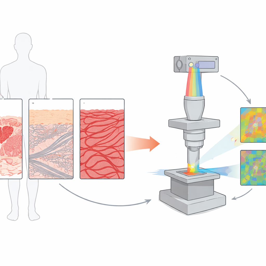
Why Light Direction Matters in Tissue
Many biological molecules interact with light differently depending on their orientation, a property known as anisotropy. Collagen fibers, for example, act a bit like rows of tiny crystals that bend polarised light in specific ways. Traditional polarised microscopes can highlight bright fibers on a dark background, but they largely provide qualitative impressions and depend heavily on the observer. The authors instead use quantitative polarisation microscopy, which measures the full polarisation state of light—how its direction and “twist” change at every pixel. From these measurements they derive maps of phase retardation (how much the light wave is delayed) and depolarisation (how much its orderly polarisation is lost), turning the invisible architecture of collagen into numbers and color-coded images.
A New Type of Polarised Microscope
To achieve this, the team built a specialised microscope using two photoelastic modulators and a lock-in detection scheme. In simple terms, they rhythmically “shake” the polarisation of the incoming light at known frequencies and synchronise the camera to those rhythms. This allows them to separate very small polarisation signals from background noise, capturing the full set of Stokes parameters that describe polarised light. From these, they compute maps of azimuth (orientation), ellipticity (how circular the polarisation has become), phase retardation, and depolarisation. Unlike standard crossed polarisers, this setup not only shows aligned fibers but also detects how disordered or complex the tissue structure is, and does so over large areas while preserving fine microscopic detail.
What Placenta, Skin, and Scars Reveal
The researchers applied this method to thin sections of human placenta, normal skin, and keloid scars, each stained either with Congo red or picrosirius red. In placenta, they found relatively low but patchy birefringence and depolarisation, with collagen forming rings around blood vessels. These subtle perivascular patterns, only weakly visible with standard cross-polarised images, were clearly captured as variations in phase retardation and depolarisation. In normal skin, especially in the deeper dermis, both measures were much stronger, reflecting thicker, more bundled collagen fibers. The superficial layers showed distinct signatures from surface keratin and underlying dermal collagen, matching known skin structure but now expressed quantitatively. Keloid scars, which are overgrown and disorganised scars, stood out even more: phase retardation rose to about 1.4 radians and depolarisation approached 0.96, indicating denser, thicker, and more chaotic collagen networks than in surrounding normal dermis.
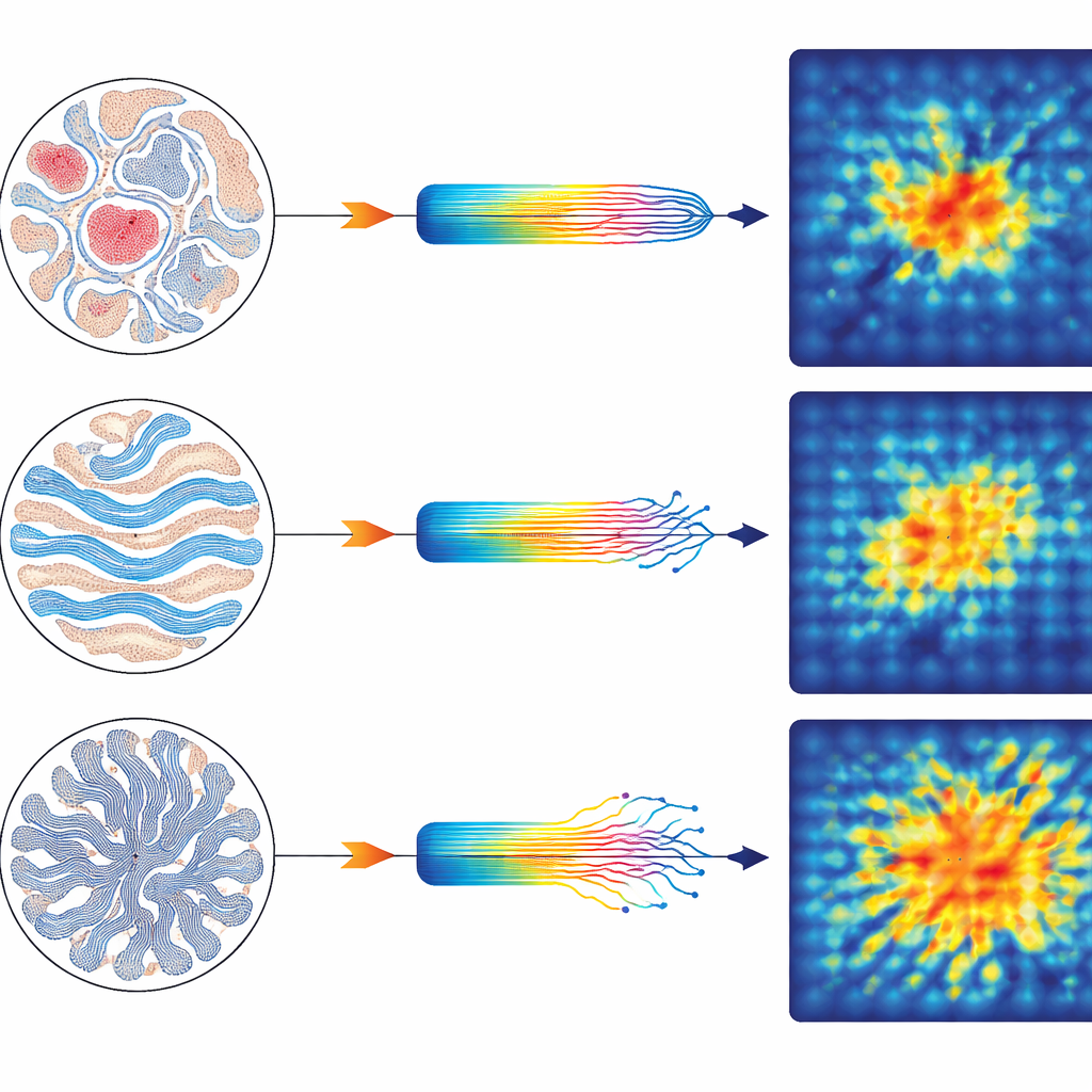
How Different Dyes Change the Picture
The team also compared two widely used histology stains that interact with collagen in different ways. Picrosirius red produced phase retardation signals three to four times higher than Congo red, confirming that it strongly boosts collagen’s birefringence by lining up along the fibrils. Congo red, by contrast, is less selective for collagen and also binds other proteins such as amyloid, leading to weaker collagen-specific enhancement. Interestingly, while picrosirius red amplified the birefringent signal, depolarisation differences between the two stains were smaller, underscoring that the stain’s chemistry mainly alters how clearly collagen’s directional effects appear, rather than the underlying tissue disorder itself.
From Research Tool to Diagnostic Aid
To a lay reader, the key message is that this technique transforms how pathologists can look at tissue. Instead of relying solely on how bright or colorful collagen appears by eye, quantitative polarisation microscopy assigns numbers to its order, thickness, and disruption. The study shows that this approach distinguishes normal from scarred skin, highlights subtle collagen patterns in placenta, and clarifies how different dyes influence what we see. Looking ahead, such measurements could help track early disease changes, guide digital image analysis and machine learning tools, and perhaps even work on unstained tissue. In essence, the authors demonstrate that carefully controlled polarised light can act as a sensitive probe of the body’s microscopic scaffolding, offering a more objective lens for understanding how tissues are built and how they break down.
Citation: Mappa, G., Miklavc, P., Cummings, M. et al. Polarimetric imaging of collagen in histopathology specimens: an investigation of congo red and picrosirius red-stained placenta and skin. Sci Rep 16, 12441 (2026). https://doi.org/10.1038/s41598-026-37711-8
Keywords: collagen imaging, polarised light microscopy, histopathology, keloid scars, picrosirius red