Clear Sky Science · en
A superior ultrafast parallel BMAS architecture for high-resolution medical ultrasound imaging
Sharper Images from Sound
Ultrasound scans are a mainstay of modern medicine, from monitoring a baby during pregnancy to checking the heart and blood vessels. Yet doctors still struggle with blurry or noisy images, especially when they need to see fine details quickly. This paper describes a new way to process ultrasound signals inside the scanner so that it produces clearer pictures at very high video-like frame rates. The work focuses on the “beamformer,” the digital core that turns raw echoes into the familiar fan-shaped ultrasound images.
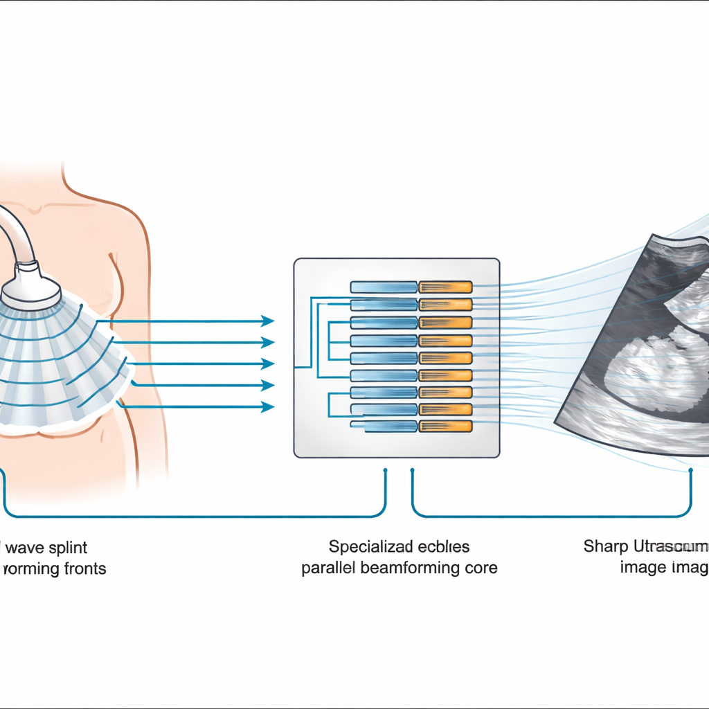
Why Current Scanners Hit a Wall
Conventional ultrasound machines use a method called delay-and-sum beamforming. In essence, echoes from dozens of tiny elements in the probe are delayed so that sound from a chosen point in the body lines up, then they are added together. This is simple and fast, but it struggles to block out stray echoes coming from other directions, which lowers contrast and makes small structures harder to see. More advanced methods that adapt to the incoming sound can improve quality, but they demand enormous computing power and energy, limiting their use in real-time clinical systems. As doctors turn to “ultrafast” imaging—capturing hundreds of frames per second for moving organs like the heart—these limits become even more pressing.
A New Way to Add Up Echoes
The authors build on a more powerful family of methods that do more than just add delayed echoes. In a technique called Beam Multiply and Sum (BMAS), coarse beams are first formed using the simple delay-and-sum approach. Instead of stopping there, these beams are then multiplied with one another in carefully chosen combinations before being added up again. This extra step emphasizes echoes that are truly coming from real structures in the body and suppresses random noise and side lobes, leading to sharper boundaries and better contrast between, for example, a fluid-filled cyst and surrounding tissue.
Splitting the Work to Go Ultrafast
Performing these extra multiplications across all 128 channels of a modern probe would normally overwhelm the digital hardware inside a scanner. To avoid this, the team designs a clever compromise. They divide the 128 channels into four smaller groups, or sub-arrays, of 32 channels each. Each sub-array first produces 28 beams in parallel using the simple method, for every broad “plane wave” pulse sent into the body. Then, for each beam direction, the four sub-array beams are combined through BMAS-style multiply-and-sum operations. This sub-array strategy slashes the number of required multipliers from thousands down to a few hundred, making the design practical for a single field-programmable gate array (FPGA) chip.
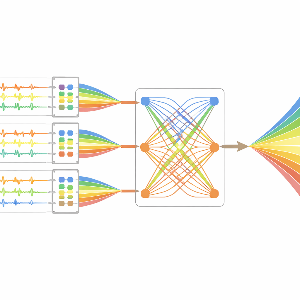
Smart Memory and Custom Hardware
To keep up with the data flow from 128 channels sampled at 40 million times per second, the researchers also redesign how timing information (delays) is stored and retrieved. They place large delay tables in external memory chips and use a special “multi-ported” delay line architecture that can read 28 different delay values at once, while writing back new ones. This arrangement, implemented with standard FPGA building blocks, allows the system to form 28 beams in parallel from each transmission without running out of on-chip memory. The entire design is coded in hardware description language and deployed on a custom ultrasound platform that includes a 128-channel transceiver board and a high-end FPGA beamformer board.
What the Images Show
The team tests their design both in simulation and with physical “phantoms,” blocks of material that mimic human tissue and contain known patterns of small cyst-like structures. They compare images from their new Parallel Beam Multiply and Sum (PBMAS) architecture with those from a conventional delay-and-sum system, using standard measures of quality such as contrast ratio and contrast-to-noise ratio. The PBMAS images show narrower beams—about 0.4 millimeters in one key measure—cleaner separation of nearby cysts, and higher contrast, indicating that subtle features should be easier for clinicians to detect. At the same time, the system sustains an impressive frame rate of 571 images per second over a 90-degree field of view, fast enough for demanding applications like heart imaging.
What This Means for Patients and Devices
In plain terms, the new architecture lets an ultrasound scanner “listen” with more intelligence while still thinking fast. By reorganizing how echoes are combined and how the hardware is used, the authors achieve clearer, higher-contrast images without giving up the ultrafast frame rates needed to watch moving organs in real time. Although the work is at the prototype stage, it shows a practical path toward future scanners that can reveal finer detail more reliably, helping doctors spot disease earlier and guide treatments more safely.
Citation: SG, S., R, S. & Kidav, J.U. A superior ultrafast parallel BMAS architecture for high-resolution medical ultrasound imaging. Sci Rep 16, 9967 (2026). https://doi.org/10.1038/s41598-026-37416-y
Keywords: ultrasound imaging, beamforming, FPGA, medical diagnostics, image resolution