Clear Sky Science · en
Deciduous pulp stem cell-derived extracellular vesicles stimulate the proliferation of cartilage progenitor cells via extracellular signal-regulated protein kinase 1/2 activation
Why baby teeth might matter for bone growth
For children born with conditions that stunt bone growth, such as certain forms of dwarfism and osteogenesis imperfecta (brittle bone disease), current treatments are limited and often only manage symptoms. This study explores an unexpected ally: cells from naturally shed baby teeth. The researchers show that tiny particles released by these dental stem cells can wake up and speed up special cartilage cells that drive bone lengthening, suggesting a future, cell-free way to help kids grow stronger, longer bones.
The hidden builders inside growing bones
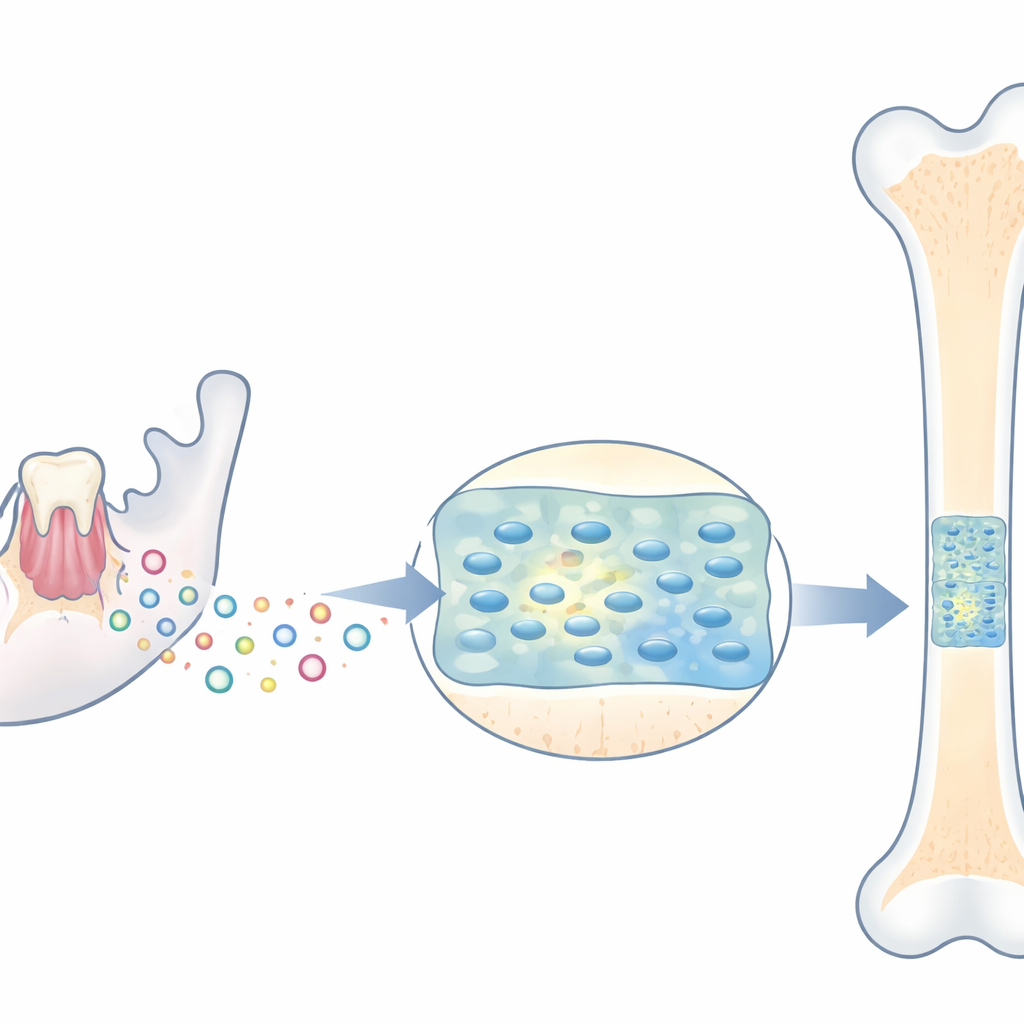
Long bones, like those in the legs and spine, lengthen at growth plates—thin zones of cartilage near their ends. Within these plates live cartilage progenitor cells, a pool of stem-like cells that divide and supply new cartilage, which later turns into bone. If these progenitor cells do not divide properly, growth slows and children can become very short in stature. The team first isolated human cartilage progenitor cells from fetal cartilage and confirmed that these cells could multiply robustly and turn into cartilage, bone, and fat cells, confirming their role as a versatile building crew for the skeleton.
Messages in microscopic packages
Stem cells from the soft pulp of discarded baby teeth continually release microscopic bubbles called extracellular vesicles. These vesicles carry proteins and other molecules that can influence distant cells, acting like parcels in a biological delivery service. The scientists purified vesicles from healthy baby-tooth stem cells and showed that cartilage progenitor cells quickly took them up. When exposed to these vesicles, the progenitor cells divided faster, moved more efficiently through the cell cycle, and showed higher activity of telomerase, an enzyme that supports long-term cell renewal and prevents premature cellular aging.
A signaling switch that drives renewal
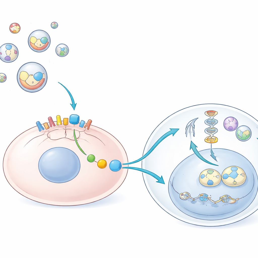
To uncover how these vesicles boost growth, the researchers focused on a signaling pathway inside cells known as ERK1/2, which acts like a molecular on–off switch for cell division. They found that the vesicles rapidly increased the activated (phosphorylated) form of ERK1/2 in cartilage progenitor cells and kept it elevated for up to an hour. Blocking ERK1/2 with a chemical inhibitor erased the benefits of the vesicles: cell division slowed, telomerase activity dropped, and the key transition between resting and DNA-copying phases of the cell cycle was impaired. This showed that ERK1/2 is essential for the vesicles’ growth-promoting effect.
How a surface handle on vesicles sparks the signal
The team then asked which component on the vesicle surface flips the ERK1/2 switch. They homed in on CD29, a membrane protein known to help cartilage cells sense their surroundings. By using gene-editing tools, they created cartilage progenitor cells lacking CD29 and also separated vesicles into CD29-rich and CD29-poor groups. Vesicles loaded with CD29 could transfer this protein onto CD29-deficient cells, restore ERK1/2 activation, and rescue their ability to divide and progress through the cell cycle. Vesicles without CD29, or blocking CD29 with specific antibodies, blunted this effect. Intriguingly, even when the vesicles were stripped of most of their RNA, they still enhanced growth, pointing to surface proteins—especially CD29—rather than genetic cargo as the main drivers.
Helping fragile bones catch up
To test relevance for disease, the researchers studied stem cells from the dental pulp of children with severe osteogenesis imperfecta, who typically have short stature and fragile bones. These patient-derived cells showed sluggish division and delayed movement through the cell cycle. Exposure to baby-tooth vesicles restored their proliferation, boosted telomerase levels, and reactivated ERK1/2 signaling. In newborn mouse tibias grown in the lab, added vesicles increased cell division and ERK1/2 activation in the growth plate, suggesting that these microscopic packages can penetrate cartilage and act where bone lengthening actually occurs.
What this could mean for future therapies
Overall, the study proposes a clear chain of events: vesicles from baby-tooth stem cells deliver the surface protein CD29 to cartilage progenitor cells, which in turn activates ERK1/2, raises telomerase activity, and speeds the crucial cell cycle steps that fuel bone growth. Because vesicles are cell-free and can be derived from discarded teeth, they may offer a safer, more practical approach than transplanting whole stem cells. While animal studies and clinical trials are still needed, this work points toward a future in which a child’s own or donated baby teeth could help treat growth plate–related disorders and improve height and bone strength without invasive procedures.
Citation: Murata, S., Sonoda, S., Kyumoto-Nakamura, Y. et al. Deciduous pulp stem cell-derived extracellular vesicles stimulate the proliferation of cartilage progenitor cells via extracellular signal-regulated protein kinase 1/2 activation. Sci Rep 16, 12654 (2026). https://doi.org/10.1038/s41598-026-37380-7
Keywords: cartilage progenitor cells, extracellular vesicles, dwarfism, osteogenesis imperfecta, dental pulp stem cells