Clear Sky Science · en
BPD-Neo: An MRI Dataset for Lung-Trachea Segmentation with Clinical Data for Neonatal Bronchopulmonary Dysplasia
Why Tiny Lungs Matter
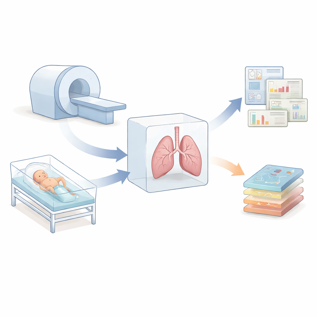
Each year, many babies are born too early, with lungs that are still under construction. These fragile lungs can lead to a serious condition called bronchopulmonary dysplasia, or BPD, which often keeps infants in intensive care for weeks or months. Doctors would like to see inside these tiny chests in three dimensions, safely and in detail, to understand what is going wrong and how to help. This article presents a new open dataset that brings together advanced MRI scans, careful outlines of the lungs and windpipe, and key medical information from 40 preterm infants, giving researchers a powerful new way to study and measure disease in the smallest patients.
From Flat X-Rays to 3D Views
Today, bedside X-rays are the workhorse for monitoring preterm infants in intensive care units, but they show only flat, two-dimensional images and expose babies to repeated radiation. Computed tomography (CT) can reveal three-dimensional structure but uses even higher doses of radiation and is rarely used in these newborns. Magnetic resonance imaging (MRI) offers a different path: it provides detailed, three-dimensional pictures of soft tissues without radiation and can be done while babies are quietly breathing and swaddled, often without sedation. In this study, the team used a motion-robust MRI technique called StarVIBE, well suited for wiggly infants, to capture high‑resolution 3D images of the lungs and the main airway.
Building a Careful Picture of Tiny Airways
The researchers scanned 40 infants younger than six months, most with some degree of BPD, using an MRI scanner located directly in the neonatal intensive care unit. To protect hearing and comfort, the babies were fed, swaddled, and given layered noise protection, while nurses closely watched vital signs. After scanning, imaging specialists used a software tool to trace out the lungs and trachea in three dimensions. They placed seed points inside each structure and let a computer algorithm grow regions outward, followed by smoothing and meticulous manual corrections. These finished outlines were saved as separate files that pair exactly with the original MRI data, and a second expert re‑segmented a subset of scans so that differences between human reviewers could be measured.
Adding Medical Details to Imaging Data
Alongside the images, the dataset includes basic but important clinical information for each infant, such as birth weight, length, sex, gestational age at birth, age at the time of scanning, and a modern grading of BPD severity using the 2019 Jensen criteria. Because every data record is fully de‑identified and linked only by a study code, families’ privacy is preserved. Having these medical details together with 3D lung and airway shapes allows researchers to explore how structural changes in the lungs relate to how early the baby was born, how much they weighed, and how severe their breathing problems became. For example, scientists can compute total lung volume, adjust it for body size, and create maps of how lung tissue signal varies, which may reflect scarring, over‑inflation, or collapsed areas.
Teaching Computers to Trace Tiny Structures
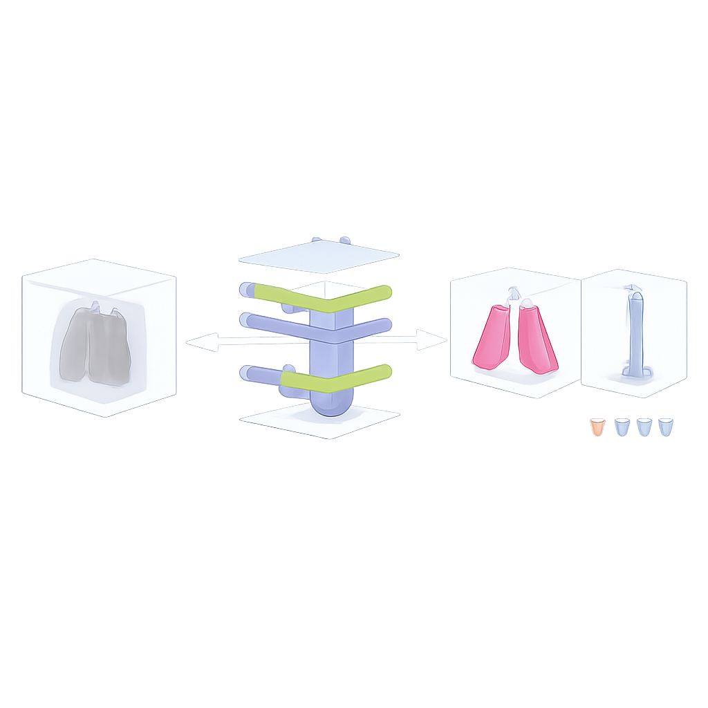
To show what can be done with this resource, the authors trained computer models known as U‑Nets to automatically find and outline lungs and tracheas in the MRI scans. These models learn from examples: they see many pairs of images and expert outlines, then try to produce similar outlines on unseen scans. Using a rigorous cross‑validation procedure, the lung model performed extremely well, reaching agreement with human experts that is close to the level two experts achieve with each other. The trachea, a much smaller and fainter structure on MRI, proved more challenging. Both humans and algorithms showed more disagreement when tracing it, and the automatic trachea outlines were less accurate and more sensitive to differences in body size and imaging conditions.
What This Means for Fragile Newborns
The BPD‑Neo dataset provides, for the first time, a freely available bundle of 3D MRI scans, expertly traced lung and airway shapes, and paired clinical data for preterm infants at risk for chronic lung disease. By showing that automated tools can already match human performance for lung volumes and flagging where current methods still struggle—such as the tiny trachea—the work lays the foundation for better, faster, and more objective measurements of lung health in newborns. In the long run, improved imaging and analysis could help doctors detect airway weakness and lung injury earlier, tailor treatments more precisely, and reduce the burden of BPD on children and their families.
Citation: Saluja, R., Kovanlikaya, A., Chien, C. et al. BPD-Neo: An MRI Dataset for Lung-Trachea Segmentation with Clinical Data for Neonatal Bronchopulmonary Dysplasia. Sci Data 13, 659 (2026). https://doi.org/10.1038/s41597-026-07006-8
Keywords: preterm infant lungs, bronchopulmonary dysplasia, neonatal MRI, lung segmentation, medical imaging dataset