Clear Sky Science · en
Super-resolution imaging of limited-size objects
Seeing the Tiny World More Clearly
Modern microscopes have allowed us to peer into cells, viruses and nanotechnology, but they still bump into a stubborn barrier known as the diffraction limit, which blurs details smaller than about half the wavelength of light. This paper shows that by using a clever way of processing light, scientists can break past that long‑standing limit without special dyes, near‑field probes or exotic tricks—opening a path to sharper views of tiny objects in physics, chemistry, materials science and even environmental monitoring.
Why Sharpness Has a Natural Limit
For more than a century, the resolution of ordinary light microscopes has been guided by the ideas of Abbe, Helmholtz and Rayleigh: no matter how perfect the lenses, details much smaller than roughly half the light’s wavelength get washed out into a blur. This is not a hard physical wall, but it is a very practical one for standard instruments. Many recent “super‑resolution” methods beat this limit, but they usually rely on fluorescent labels, near‑field scanning, or structured illumination that complicate experiments and can disturb delicate samples. The authors revisit the problem from the standpoint of information theory, treating an imaging system as a channel that carries information from object to detector, and ask: how much detail can we recover if we only assume that the object fits within a small region of space?
A New Way to Use What We Already Know
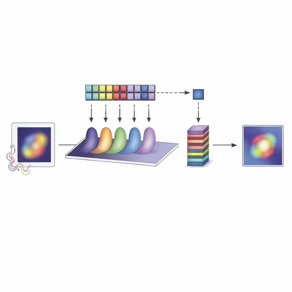
The central idea is surprisingly simple: if you know that everything you care about lies inside a tiny patch—smaller than a wavelength of light—it becomes possible, in principle, to reconstruct features much finer than the diffraction limit. The team builds on a mathematical family of functions called Slepian–Pollak modes, which efficiently describe any pattern confined to a limited field of view. Instead of trying to directly form an image, their method, called limited‑size object microscopy (LSOM), measures how strongly the scattered light from the object excites each of these modes. By carefully recovering a finite set of mode “weights,” they can rebuild the near‑field pattern of light around the object with far higher detail than conventional imaging would suggest.
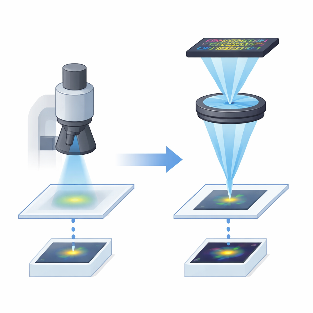
Turning Blurred Light into a Sharp Picture
To make this work in the lab, the researchers designed a microscope that treats each Slepian–Pollak mode almost like a separate communication channel. A nanoparticle on a sapphire cube is illuminated so that it scatters coherent light, which is collected by a high‑quality objective lens. In the plane where the lens focuses different angles of light, a programmable digital micromirror device acts as a reconfigurable mask that can pick out one mode at a time and interfere it with a strong reference mode. By cycling through tailored mask patterns and recording the resulting light with a camera pixel that acts as a sensitive single‑pixel detector, the system measures both the strength and phase of each mode. A carefully calibrated mathematical filter then compensates for imperfections in the optics and converts these measurements into accurate mode coefficients.
Beating the Diffraction Limit in Practice
Armed with this setup, the team imaged platinum and gold nanoparticles with various shapes and sizes, all confined to regions smaller than 0.8 times the wavelength used. They reconstructed images using only a modest number of modes—13 for two‑dimensional shapes and 6 for one‑dimensional lines—yet achieved effective resolutions as fine as one‑seventh to one‑eighth of the wavelength, well beyond the usual diffraction limit. Independent checks confirmed this performance: the point‑spread function of the system was several times narrower than that of a standard microscope, a nanoscale “Siemens star” test pattern showed clearly separated features at λ/7 spacing, and the recovered mode coefficients closely matched numerical simulations even for weak, high‑order modes.
What This Breakthrough Means
The study demonstrates that deep super‑resolution imaging is possible in the far field, without labels or structured illumination, if we exploit a simple piece of prior knowledge: that the object occupies a limited area. For isolated nano‑objects—such as engineered nanoparticles, nanowires, or tiny pollutants in air or water—this is often a very natural assumption. Because LSOM can be implemented by adding a relay and programmable mask to a conventional microscope, it offers a practical route to sharper imaging in many laboratories. Beyond microscopy, the same approach to recovering finely detailed light patterns could improve precision measurements in metrology, spectroscopy and optical ranging, helping scientists and engineers see and measure the nanoscale world with unprecedented clarity.
Citation: Chang, T., Adamo, G. & Zheludev, N.I. Super-resolution imaging of limited-size objects. Nat. Photon. 20, 421–427 (2026). https://doi.org/10.1038/s41566-025-01839-2
Keywords: super-resolution imaging, label-free microscopy, nano-objects, optical diffraction limit, photonics