Clear Sky Science · en
Fructose-1,6-bisphosphate couples glycolytic activity to cell adhesion
How Sugar Talks to a Cell’s Grip
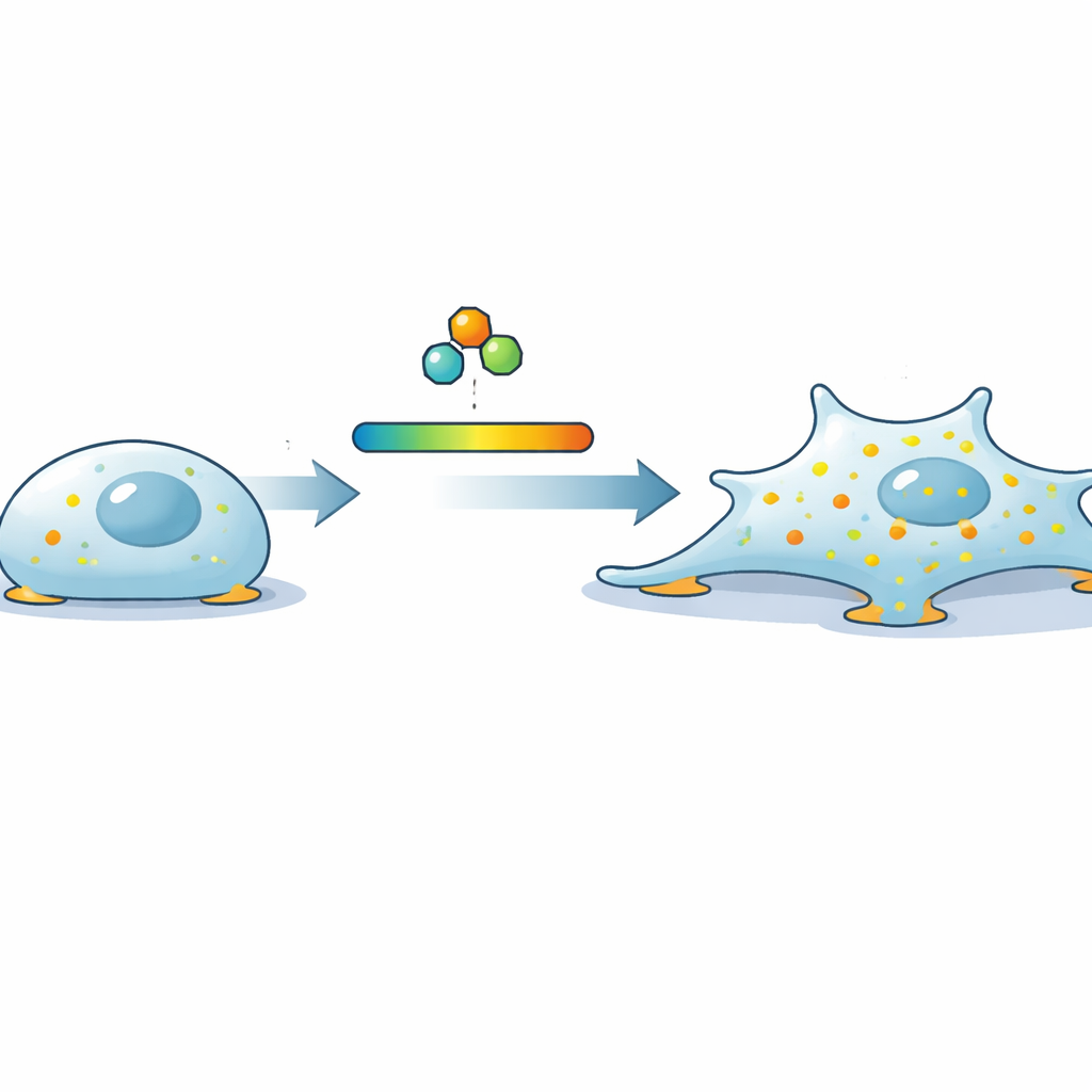
Every time a cell crawls across a surface, heals a wound, or begins to spread as a tumor, it must decide how tightly to hold on and how boldly to push forward. This study reveals that a familiar sugar breakdown product inside cells, called fructose-1,6-bisphosphate (FBP), acts like a molecular dimmer switch that links how much sugar the cell burns to how strongly it sticks and spreads. Understanding this hidden conversation between metabolism and cell adhesion could reshape how we think about development, immunity, and cancer invasion.
Discovering a Hidden Connection
To uncover what controls the tiny anchoring sites that cells use to grip their surroundings, the researchers performed a massive gene-silencing screen. They used automated microscopy to inspect more than 18,000 genes in human cells, looking for changes in “focal adhesions,” the small, dot-like structures that connect the cell’s internal scaffolding to the outside world. Among the strongest hits was an unexpected player: aldolase A, a classic enzyme of glycolysis, the pathway that breaks down glucose for energy. When aldolase A was reduced, cells formed many more focal adhesions and spread out over a larger area, a change that was reversed when a normal version of the enzyme was put back.
Sugar Metabolite as a Signal, Not Just Fuel
At first glance, it would be easy to assume that this effect was simply about energy: alter glycolysis, change ATP levels, and cells behave differently. But the story turned out to be more subtle. When the team knocked down other glycolytic enzymes, all conditions lowered ATP, yet only manipulating the steps that control FBP levels changed adhesion. Reducing the enzyme PFK, which produces FBP, caused cells to shrink and lose adhesions, the opposite of aldolase loss. In contrast, removing an enzyme further downstream had little effect on cell shape or grip. Direct measurements showed that high FBP levels tracked closely with large, strongly adherent cells, while low FBP went with small, weakly attached ones. Blocking FBP production, either genetically or with a glycolysis inhibitor, could “reset” aldolase-deficient cells back to normal, proving that it was the concentration of this one metabolite—not energy supply overall—that was steering adhesion.
From Inner Chemistry to Outer Shape
How does a small metabolite re-sculpt the cell’s outer edge? Using live-cell imaging of focal adhesion markers at the cell’s underside, the scientists found that FBP boosts the birth of new adhesions rather than slowing their breakdown. High FBP increased the rate at which adhesions assembled and the number of new sites formed, while low FBP had the opposite effect. At the same time, the actin cytoskeleton—the dynamic network of protein fibers that drives movement—was reorganized. Cells rich in FBP displayed broad, sheet-like protrusions and more complex actin patterns, and they spread faster across surfaces, whereas FBP-poor cells showed fewer stress fibers and less protrusive edge. Importantly, naturally occurring situations that demand active spreading and migration, such as cells reseeding or moving, were accompanied by strong rises in FBP to levels similar to those seen in the experimental manipulations.
A Molecular Brake Is Released
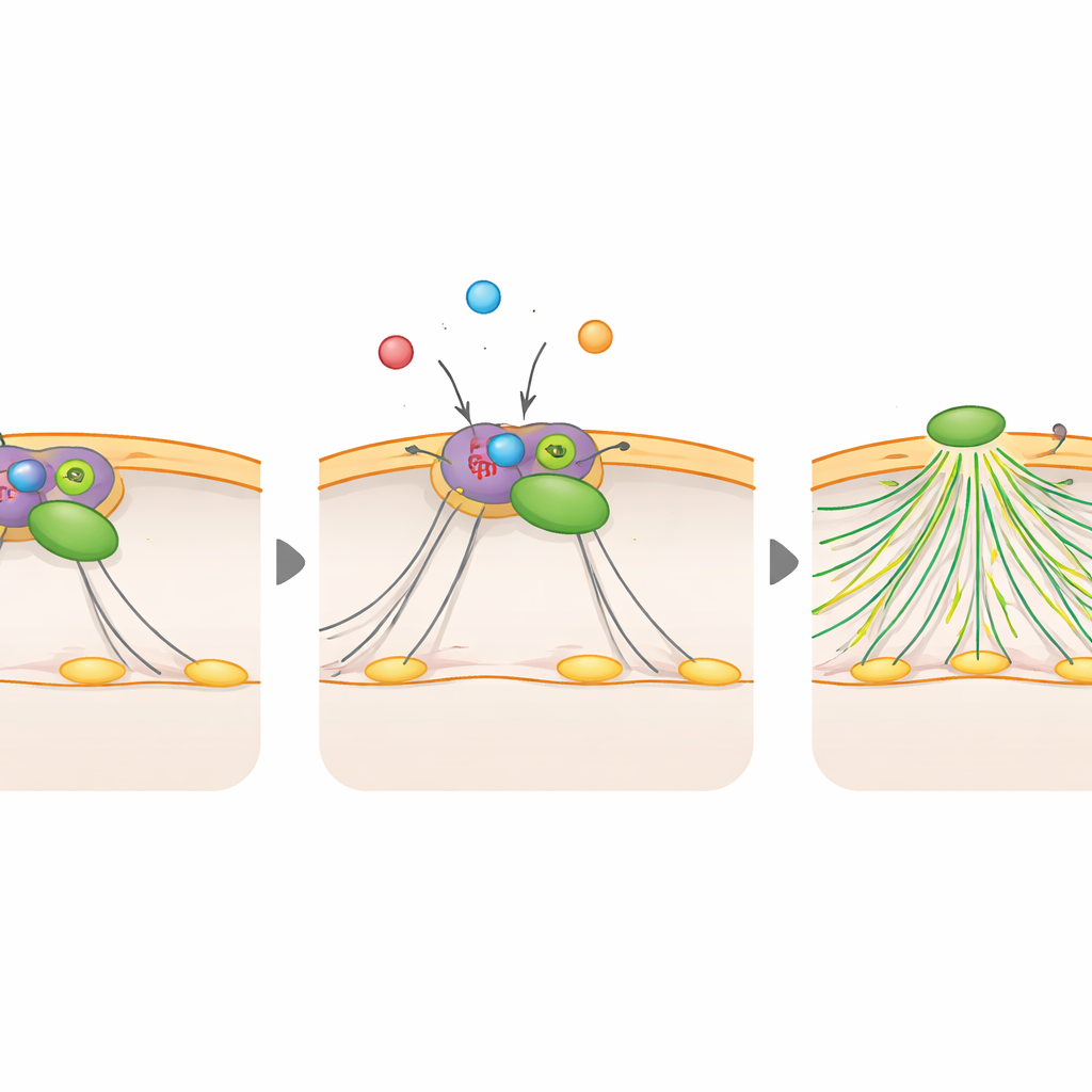
To connect this behavior to known control circuits, the team searched for proteins that change shape when exposed to FBP inside cell extracts. This search pointed to Rac1, a master regulator of actin-driven protrusions, and RCC2, a protein that can hold Rac1 in an inactive complex. The researchers showed that high FBP levels increase Rac1 activity, while low FBP dampen it. When Rac1 was removed or forced into an inactive form, the extra adhesions and spreading caused by high FBP disappeared; when Rac1 was locked in an active state, it could bypass the effects of low FBP. Biochemical tests revealed that FBP binds directly to RCC2 and weakens its interaction with Rac1. In essence, FBP pries Rac1 loose from its inhibitor, allowing other factors to switch Rac1 on, which then triggers actin remodeling, new protrusions, and more adhesion sites.
Why This Matters for Health and Disease
This work shows that FBP serves as more than a stepping stone in energy production—it is also a messenger that lets cells sense when glycolysis is running high and adjust their physical behavior accordingly. When sugar breakdown intensifies, FBP rises, frees Rac1 from RCC2, and encourages cells to spread and explore; when glycolysis is low, Rac1 remains restrained and cells stay compact and less adhesive. Such a mechanism likely influences early development, where glycolysis is prominent, as well as cancer, immune cell movement, and blood vessel growth, all of which rely on both high glycolytic activity and dynamic cell protrusions. By revealing how a simple metabolic intermediate tunes the cell’s grip on its surroundings, this study highlights a direct chemical link between what a cell eats and how it moves.
Citation: Hoffmann, L., Duchmann, M., Lazarow, K. et al. Fructose-1,6-bisphosphate couples glycolytic activity to cell adhesion. Nat Cell Biol 28, 739–753 (2026). https://doi.org/10.1038/s41556-026-01911-1
Keywords: cell adhesion, glycolysis, Rac1 signaling, cell migration, cancer invasion