Clear Sky Science · en
Mathematical modeling of planar cell polarity: principles, approaches, and open questions
How cells agree on a shared direction
Many organs in our bodies, from the inner ear to the airways and kidneys, rely on sheets of cells that all "face" the same way across the tissue surface. This coordinated alignment, called planar cell polarity, acts like a built‑in compass that guides how tissues grow, bend, and function. When this compass goes wrong, the consequences can be serious, contributing to birth defects, hearing loss, respiratory problems, and spine deformities. This article reviews how scientists use mathematical and computational models to understand how individual molecules inside cells team up to create such large‑scale order.
Why tissue-wide direction matters
Planar cell polarity (PCP) describes how cells in a thin sheet align in a common direction, sideways across the tissue rather than top‑to‑bottom. In fruit flies, PCP first attracted attention when tiny wing hairs all pointed in the same direction. Similar directional patterns appear in vertebrates: nerve tissues close correctly only if cells extend and intercalate in aligned ways; hair cells in the inner ear must be oriented precisely to detect sound; cilia in the airways and brain ventricles must beat in sync to move fluid; and hair follicles in the skin follow coordinated patterns. Disruptions in the genes that control PCP are linked to neural tube defects, skeletal disorders such as Robinow syndrome, kidney malformations, and scoliosis. Because PCP shapes organs in both embryos and adults, researchers want to know how molecules inside each cell combine local information with long‑range cues to keep tissues properly aligned.
Two molecular compass systems
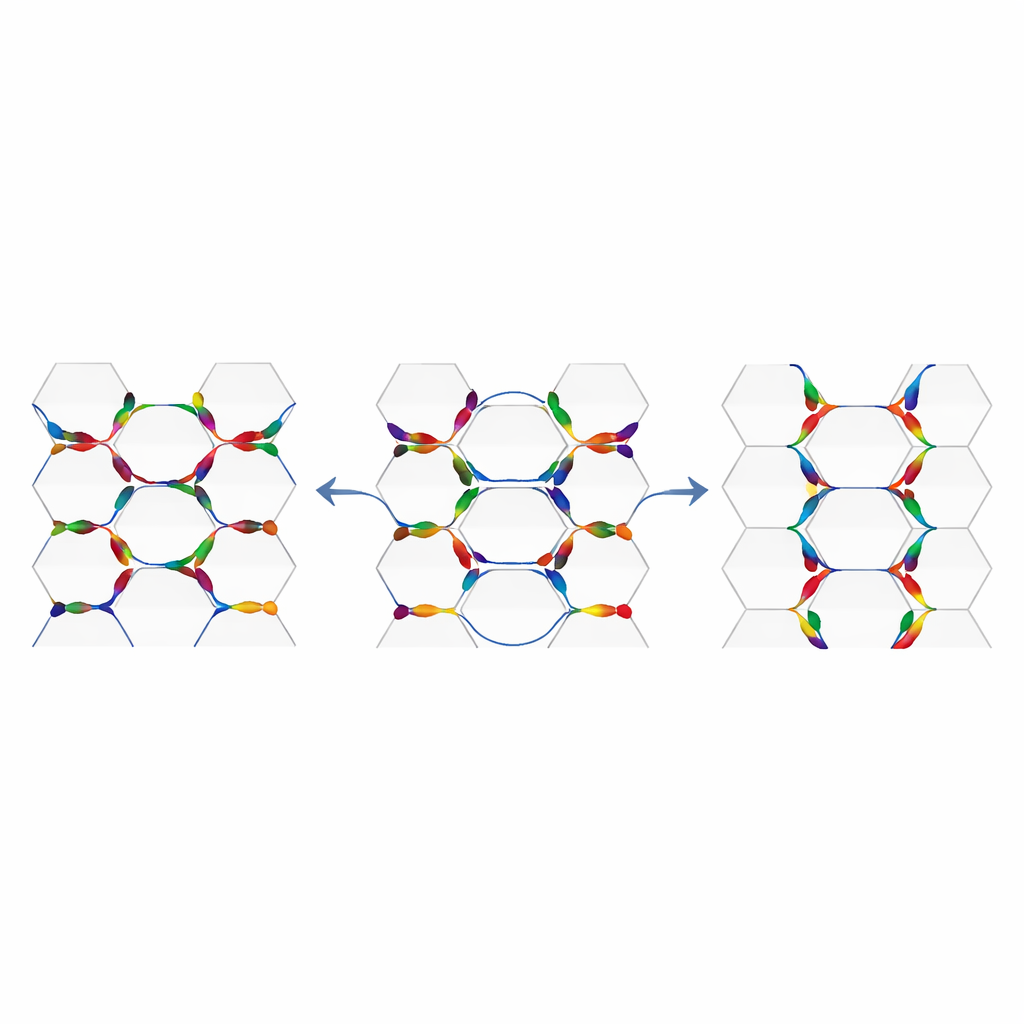
Biologists have identified two interlinked sets of proteins that act together to build PCP. One, known as the core module, contains membrane and cytoplasmic proteins that cluster asymmetrically on opposite sides of each cell. These clusters on neighboring cells interact across cell junctions, allowing cells to compare their orientation with their neighbors. A second, more global module is built from large cadherin proteins called Fat and Dachsous, plus a modifying enzyme called Four‑jointed. These components are expressed in tissue‑scale gradients, so that one side of a tissue sees slightly more of one protein than the other. That gradient subtly biases where complexes form on each cell, helping to align the local core machinery with the overall axis of the organ. Exactly how these two modules talk to each other—whether one feeds into the other in sequence or whether they run in parallel—remains an active area of investigation.
Different modeling lenses on the same problem
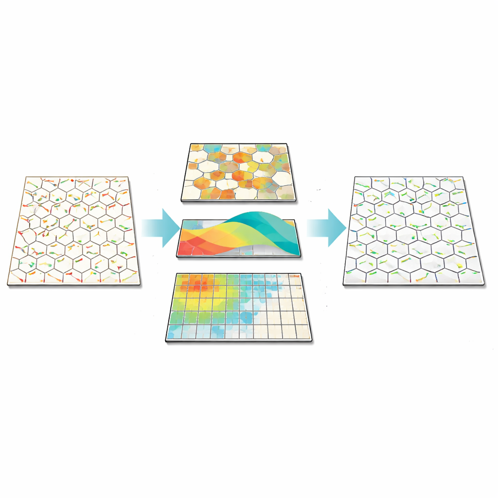
The review explains how several families of mathematical models, each with its own strengths, have been built to explore PCP. Cellular Potts models represent tissues on a grid, tracking detailed cell shapes and the positions of polarity proteins along cell edges; they show how transient or localized cues can be amplified into long‑range order. Petri net models focus on discrete binding and unbinding events of proteins in small membrane regions, capturing the stochastic nature of molecular encounters. Agent‑based models treat each cell as an entity whose membrane protein levels and interactions change over time according to reaction‑like rules; these models are widely used to study how feedback, noise, gradients, and cell geometry combine to produce tissue‑scale patterns. Phenomenological models compress all molecular detail into simple polarity vectors and energy‑like functions, borrowing ideas from magnetism to analyze when ordered patterns appear or break down. Finally, continuum theories smooth over individual cells to describe polarity as a continuous field obeying differential equations, enabling analytical insight into large tissues and links to other pattern‑forming systems.
What the models are teaching us
Across these approaches, several common lessons emerge. Local feedback between neighboring junctions can generate asymmetry even from weak global cues, but gradients of proteins such as Fat and Dachsous help align polarity across large domains and make patterns more resistant to random fluctuations. Cell‑intrinsic processes, such as how proteins move and interact inside a cell, become especially important when external signals are noisy or weak. Cell shape and tissue geometry also matter: when cells are elongated or tissues are stretched, polarity tends to align in predictable directions relative to that deformation, hinting at deep links between mechanics and molecular signaling. Models show how random molecular noise or fixed irregularities in protein levels can create swirling or patchy polarity patterns, and how clones of mutant cells can disturb the orientation of their neighbors—a behavior known as domineering non‑autonomy that is seen experimentally. At the same time, many distinct assumptions about the microscopic details can reproduce similar large‑scale behavior, underscoring how hard it is to infer the exact molecular rules from tissue‑level observations alone.
Open puzzles and future directions
Despite major progress, modeling highlights several unresolved questions. Do the two PCP modules usually work in series or in parallel, and does the answer change from one tissue to another? Is PCP best thought of as an equilibrium process, like magnets aligning in a field, or as a driven, out‑of‑equilibrium system more akin to flocking birds? How strongly do tissue flows, mechanical forces, and organ shape feed back on molecular polarity, and can unified models connect these elements across scales? The authors argue that answering these questions will require tight collaboration between theory and experiment, guided by models that make sharp, measurable predictions.
What this means for health and disease
For non‑specialists, the key message is that the orderly architecture of our organs depends on countless microscopic decisions made by proteins at cell borders, and that mathematics is becoming essential for decoding how those decisions add up. By comparing different modeling frameworks and the insights they provide, this review sketches a roadmap for building predictive theories of planar cell polarity. Such theories could ultimately help explain why certain birth defects arise, why some tissues are more robust to damage than others, and how we might one day steer tissue organization in regenerative medicine or disease.
Citation: Rizvi, M.S., Jolly, M.K. Mathematical modeling of planar cell polarity: principles, approaches, and open questions. npj Syst Biol Appl 12, 56 (2026). https://doi.org/10.1038/s41540-026-00679-2
Keywords: planar cell polarity, epithelial tissue, mathematical modeling, tissue morphogenesis, cell signaling