Clear Sky Science · en
Paired DNA and RNA sequencing uncovers common and rare variation regulating human retinal gene expression
Why tiny changes in eye DNA matter
Why do some people lose their sight as they age while others keep sharp vision, even when they share the same diagnosis or risk factors? This study looks inside donated human eyes to link small differences in DNA to how retinal cells switch genes on and off. By pairing complete DNA sequencing with detailed snapshots of gene activity, the researchers create a reference map that can help explain both common eye diseases, like age-related macular degeneration, and rare inherited blindness.
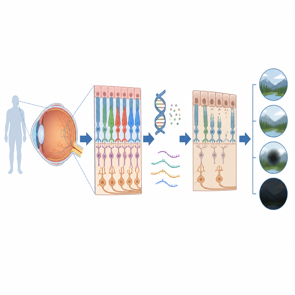
Looking closely at the back of the eye
The retina is the light-sensing film at the back of the eye, and it relies on a tight partnership between two layers. One, called the neurosensory retina, contains the nerve cells that capture light and send signals to the brain. The other, the retinal pigment epithelium, supports and nourishes those cells. The team obtained 201 post-mortem eyes from donors without late-stage macular disease or known genetic eye syndromes. From each eye they extracted DNA and also measured which genes were active in the two retinal layers, capturing tens of thousands of genes and their activity levels with very deep RNA sequencing.
Finding common DNA changes that tune retinal genes
Using these data, the scientists searched for common DNA variants that consistently raised or lowered the activity of nearby genes, a type of signal known as an expression quantitative trait locus. They discovered almost 1.5 million such links across the two retinal layers, affecting more than ten thousand genes. Many of these connections matched results from earlier retina studies, which builds confidence in the new resource. Others were brand new, including hundreds of genes not previously known to be under genetic control in the eye, and several connected to known risk regions for age-related macular degeneration. The variants were especially likely to fall in stretches of DNA that act as on–off switches or volume knobs for genes, particularly those active in retinal cell types such as rods and cones.
How disease genes behave differently
The researchers next compared genes already tied to rare inherited eye disorders with genes not yet linked to eye disease. They found that disease genes are, on average, more strongly expressed and show less natural fluctuation in activity between people. These genes also tended to have fewer common tuning variants, and when such variants were present, their effects on gene activity were smaller. In contrast, genes not associated with eye disease showed more numerous and stronger tuning variants. This pattern suggests that the retina may tolerate only modest shifts in the activity of crucial vision genes, while being more relaxed about variation in other genes.
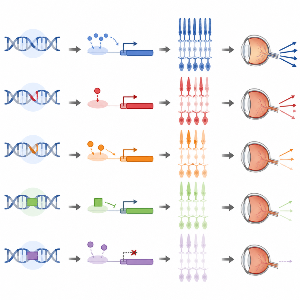
Rare disruptions that stand out from the crowd
Beyond common variants, the team hunted for rare changes that cause a gene’s activity in a given person to be unusually high or low compared with all other samples. They detected just over a thousand such outlier events and then used two computational strategies to search the surrounding DNA for likely culprits. About half of the outliers could be linked to rare mutations that either broke the gene’s protein-coding sequence or altered nearby control regions, including structural changes that delete or duplicate chunks of DNA. One striking example was a rare change in the promoter, a control patch upstream of a gene called CAND2, which has been associated with macular degeneration risk. A lab test confirmed that this single letter change weakened the promoter’s ability to drive gene activity, demonstrating that the prioritization approach can pinpoint functionally important variants.
What this means for future eye care
For non-specialists, the key message is that the health of the retina depends not only on which genes people carry but also on how strongly those genes are turned on or off in specific eye tissues. This study delivers a detailed map linking DNA variation, both common and rare, to gene activity in the two main retinal layers that support vision. Clinicians and researchers can now use this map to better interpret genetic test results, especially changes in non-coding DNA that do not alter proteins directly but can still disturb how the retina works. Over time, this kind of resource may help explain why eye disease risk and severity differ so widely between individuals and guide the search for new strategies to protect sight.
Citation: Sampson, J., Segrè, A.V., Bujakowska, K.M. et al. Paired DNA and RNA sequencing uncovers common and rare variation regulating human retinal gene expression. Nat Commun 17, 4595 (2026). https://doi.org/10.1038/s41467-026-72979-4
Keywords: retina, gene expression, age related macular degeneration, genetic variation, eye disease