Clear Sky Science · en
150 MHz polymer resonator for optoacoustic mesoscopy based on a tapered optical fiber
Seeing Tiny Blood Vessels with Sound and Light
Doctors and researchers increasingly rely on images of very small blood vessels just under the skin to study diseases, guide treatments, and track healing. A technique called optoacoustic mesoscopy combines light and ultrasound to reveal this hidden micro‑world, but it demands tiny, very sensitive ultrasound detectors. This paper presents a new hair‑thin sensor built on the tip of an optical fiber that can pick up extremely high‑frequency sound waves, allowing clearer, sharper views of fine structures such as capillaries in living tissue.
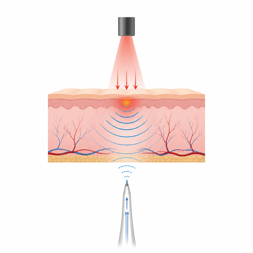
How Light Turns into Sound for Imaging
In optoacoustic imaging, very short laser pulses shine onto tissue and are absorbed by components like blood. This brief heating causes the tissue to expand slightly, launching ultrasound waves that travel outward. By recording these waves from many positions and frequencies, a computer can reconstruct a three‑dimensional picture of the structures that produced them. To see very small features, like vessels thinner than a human hair, the system must detect ultrasound over a broad range of high frequencies, up to and beyond 100 MHz, which is far higher than in conventional medical ultrasound.
Limits of Today’s Tiny Microphones
Existing miniature ultrasound detectors face tough trade‑offs. Traditional piezoelectric devices lose sensitivity as they shrink and struggle to cover very high frequencies. Optical detectors on silicon chips can be extremely small and fast, but their stiff materials reflect sound poorly and generate surface acoustic waves that skim along the surface and blur images. Polymer‑based detectors couple better to sound and avoid many of these artifacts, yet they have been difficult to miniaturize without losing optical performance, which has kept their useful frequency range relatively low and limited the image resolution they can provide.
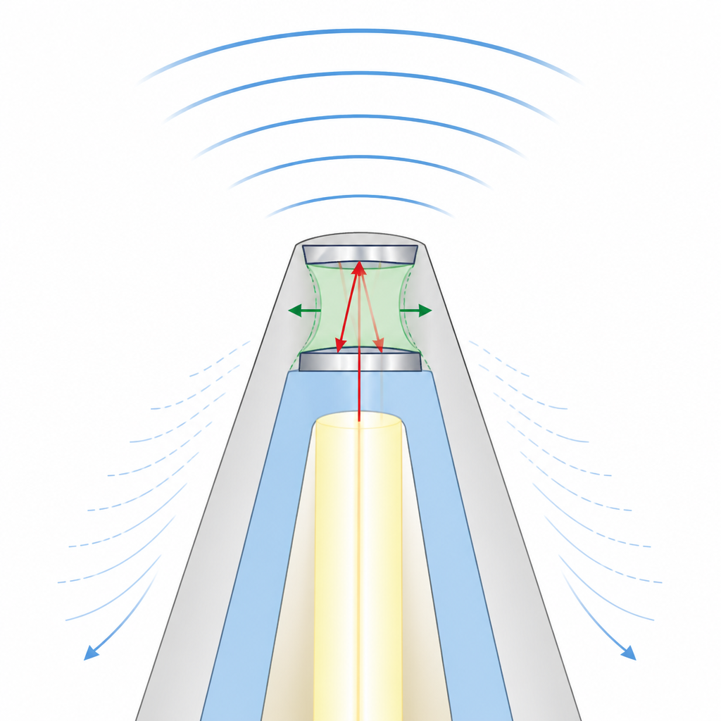
A New Fiber‑Tip Sensor Design
The authors introduce a different approach: a tiny polymer “echo chamber” built on the flattened tip of a tapered optical fiber. The fiber is polished into a cone so that only a small plateau remains at the end, and this plateau hosts a micrometer‑scale cavity made of transparent polymer sandwiched between thin silver mirrors. Light is sent down the fiber and bounces inside this cavity. When an incoming ultrasound wave slightly squeezes or stretches the polymer, the distance between the mirrors changes, altering the reflected light in a way that can be measured. By carefully shrinking both the cavity thickness and its diameter, the researchers achieved a smooth, ultra‑broad frequency response around 150 MHz, while the small active area reduced unwanted surface waves and directional bias.
Sharper Images of Tiny Vessels
The team built three versions of the sensor with different sizes to study how miniaturization affects performance. The smallest, with a base only 24 micrometers wide and a 6 micrometer‑thick polymer cavity, delivered the best results: a bandwidth of about 150 MHz and a noise‑equivalent pressure density of roughly 1.5 milli‑Pascal per square‑root Hertz, indicating very high sensitivity. Its tiny aperture gave an almost point‑like response, reducing blurring and artifacts that plagued larger designs. When used in optoacoustic mesoscopy experiments on mouse ears, the sensor produced three‑dimensional images that resolved vessels around 17–20 micrometers in diameter, with axial resolution of about 7 micrometers and lateral resolution near 17 micrometers. Frequency‑based color views highlighted smaller and larger vessels separately, revealing fine details in the skin’s microvasculature.
Toward Compact Probes and Endoscopes
Because the new detector is built on a standard optical fiber with a polymer cavity formed in a simple wet‑lab process, it avoids the need for complex chip fabrication and can be produced more easily and cheaply. The authors also show that the same tapered‑fiber concept can be extended to multi‑core fibers that both deliver light and detect sound, hinting at compact probes for endoscopy or other space‑limited settings. In plain terms, this work demonstrates a very small, very sensitive optical “microphone” for ultrasound that combines high clarity, broad frequency coverage, and fewer image artifacts, opening the door to clearer images of tiny structures inside the body.
Citation: Ülgen, O., La, T.A., Zakian, C. et al. 150 MHz polymer resonator for optoacoustic mesoscopy based on a tapered optical fiber. Nat Commun 17, 4328 (2026). https://doi.org/10.1038/s41467-026-72815-9
Keywords: optoacoustic imaging, ultrasound sensor, optical fiber, microvasculature, polymer resonator