Clear Sky Science · en
A comparative transcriptomic analysis of mouse demyelination models and multiple sclerosis lesions
Why this research matters
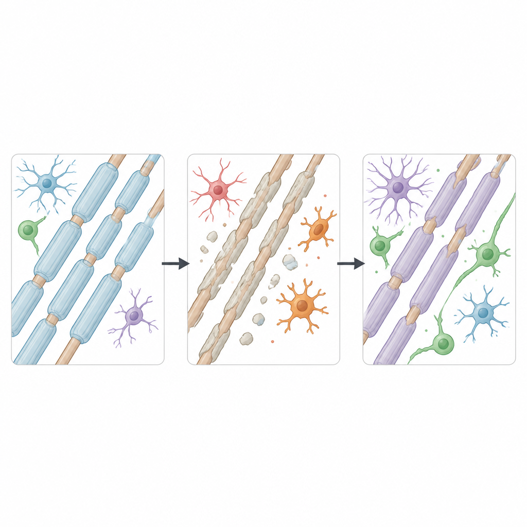
Multiple sclerosis is a disease in which the protective coating around nerve fibers, called myelin, breaks down. Scientists often rely on mouse models to study how myelin is lost and rebuilt, and to test new treatments. But not all models mimic the human disease in the same way. This study asks a practical question that matters for every future therapy: which mouse models best capture the changes seen in the brains of people with multiple sclerosis, and for which cell types?

Two ways to damage myelin in mice
The researchers focused on two widely used mouse methods that strip away myelin in the brain. One uses a chemical called cuprizone mixed into the animals' food, causing widespread loss of myelin-making cells over several weeks. The other injects a detergent-like substance, lysophosphatidylcholine, into a small brain region, creating a focused injury that heals over about a month. Both approaches reliably cause demyelination followed by regrowth of myelin, but until now it was unclear whether they trigger similar or very different responses inside brain cells, or how well either one copies what happens in human multiple sclerosis lesions.
Reading the activity of thousands of single cells
To answer this, the team used single-cell and single-nucleus RNA sequencing, a method that reads which genes are switched on in individual cells. They combined their new mouse data with earlier datasets and compared them to a large collection of human white matter samples from people with multiple sclerosis and from controls. These human samples covered many lesion types, including areas that look normal, actively inflamed regions, long-standing scars, and spots where myelin has regrown. By mapping gene activity across hundreds of thousands of cells, they built a detailed atlas of how different brain cell types respond to myelin loss and repair in mice and in humans.
Stressed and immune-like myelin cells
One key focus was oligodendrocytes, the cells that build and maintain myelin. The study uncovered a striking difference between the two mouse models. Cuprizone caused oligodendrocytes to enter a highly stressed state marked by genes linked to DNA damage, protein-folding stress, and a shift toward a senescence-like condition. This state closely resembled patterns seen in human multiple sclerosis lesions, including the activation of genes such as CDKN1A and NUPR1 that are tied to cell cycle arrest and resistance to a type of iron-driven cell death. In contrast, the detergent model did not show this same depth of stress response, suggesting it is less suited for probing how oligodendrocytes fail or become dysfunctional in chronic disease.
Despite their differences during active damage, both mouse models converged on a similar oligodendrocyte state during remyelination. In this shared state, newly formed myelin cells turned on many immune-related genes, including those involved in responding to interferons and displaying protein fragments to the immune system. A comparable immune-colored state was also present in human lesions. This suggests that myelin-forming cells themselves can adopt an “alert” profile after injury, potentially influencing inflammation and repair, not just passively rebuilding insulation.
Immune cells in the brain show shared and unique patterns
The team also examined microglia and related immune cells that patrol the brain and help clear myelin debris. In both mouse models, these cells shifted from a calm, house-keeping identity into a damage-associated form geared toward engulfing debris, handling fats, and producing inflammatory signals. Many of the same genes were switched on in microglia from human multiple sclerosis lesions, indicating a conserved core response. However, the detergent model drove a more intense and long-lasting inflammatory profile, with extra involvement of blood-derived macrophages, while cuprizone produced a briefer, more diffuse reaction. Human lesions showed even richer diversity, with distinct microglial states linked to active inflammation, chronic damage, or regions undergoing repair, reflecting the long, uneven course of the disease in people.
What this means for future studies
Overall, the study shows that no single mouse model fully reproduces the complexity of multiple sclerosis, but each captures specific pieces of the puzzle. Cuprizone best mirrors the deep stress and partial failure of myelin-forming cells seen in patients, making it useful for studying how these cells are damaged and how they might be rescued. The detergent model better represents intense, localized inflammation and prolonged activation of brain immune cells, which is helpful for probing how immune activity shapes myelin repair. By laying out where the models match or diverge from human disease, this work offers a practical guide for choosing the right experimental system to tackle particular questions about myelin injury, inflammation, and the chances for lasting repair.
Citation: Aboelnour, E.L., Vanoverbeke, V.R., Maupin, E.A. et al. A comparative transcriptomic analysis of mouse demyelination models and multiple sclerosis lesions. Nat Commun 17, 3858 (2026). https://doi.org/10.1038/s41467-026-72383-y
Keywords: multiple sclerosis, demyelination, oligodendrocytes, microglia, remyelination