Clear Sky Science · en
Reticulate leaf venation in Pilea peperomioides is a Voronoi diagram
How a houseplant hides a math puzzle
Many people grow the Chinese money plant for its round, coin-like leaves, but few would guess that those leaves quietly follow a rule from geometry class. This study shows that the largest veins in these leaves arrange themselves much like a classic puzzle called a Voronoi diagram, a way of dividing space into regions around special points. By revealing both the pattern and a step-by-step biological mechanism that can create it, the work links everyday plant shapes to simple mathematical rules.
Seeing patterns in leaf veins
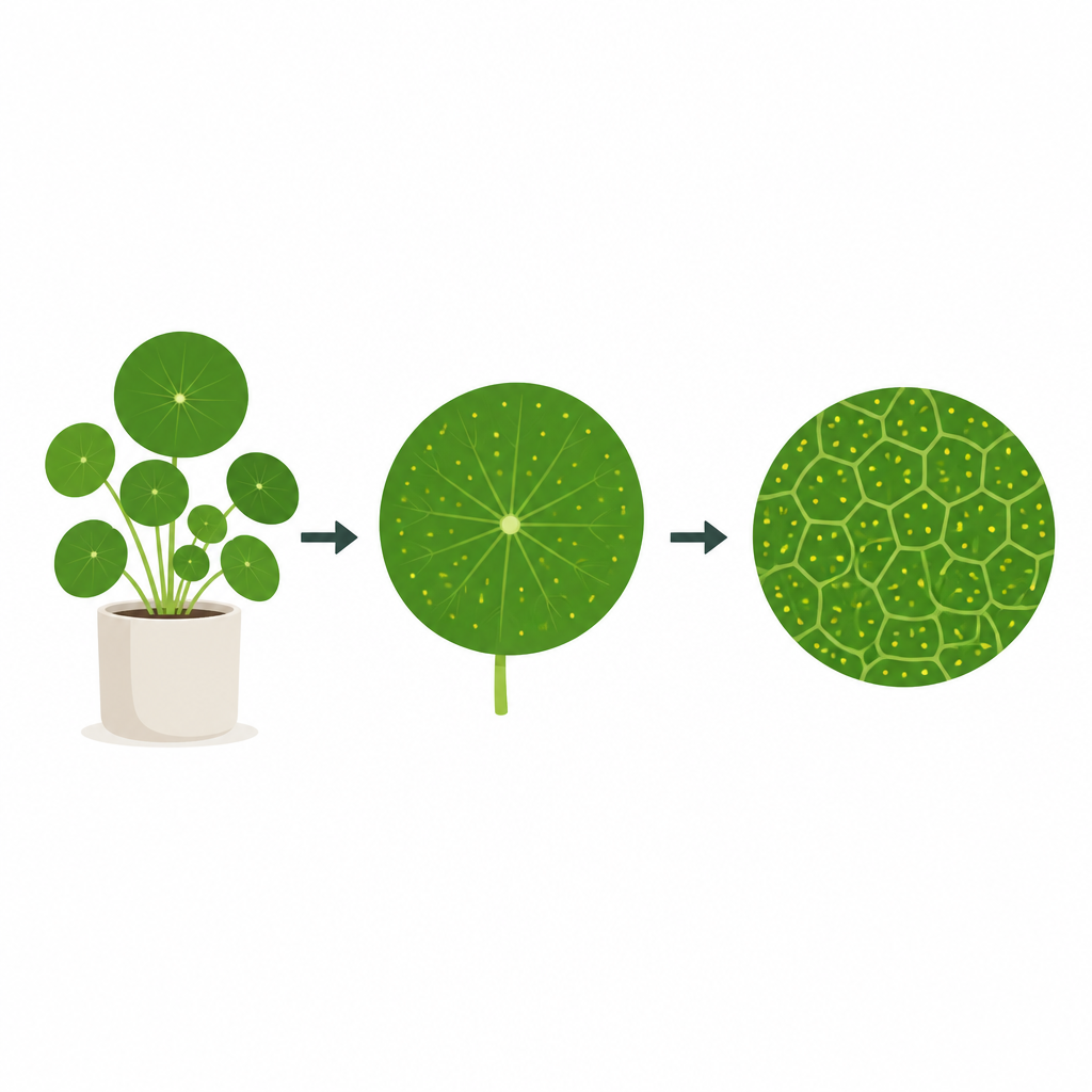
The authors focus on Pilea peperomioides, whose nearly circular leaves are attached to the stem by a stalk from below. Each leaf contains a network of thick "major" veins that form closed loops and a finer mesh of small veins. Scattered across the leaf surface are hydathodes, tiny pores that release water and help manage the leaf’s internal balance. When the researchers stained and imaged flattened leaves, then traced every major vein and every hydathode with computer assistance, they noticed a striking fact: most of the smallest loops of major veins enclosed exactly one hydathode. This suggested that the veins might act like borders drawn halfway between neighboring pores.
To test this idea, they turned to Voronoi diagrams, which partition space into cells around a set of points so that every location belongs to the nearest point. The researchers compared the real vein loops to ideal Voronoi cells built from the hydathode positions. They used three independent geometric tests: one checked whether lines between neighboring hydathodes met the shared vein boundary at right angles and equal distances; another measured how much area each real loop shared with its matching Voronoi cell; a third worked backwards, asking where the best-fitting Voronoi centers should lie for the observed network and how close those centers were to the actual hydathodes. Across all tests, hydathodes consistently behaved more like Voronoi centers than several alternative reference points inside each loop.
Patterns that persist under stress
Biological growth is never perfectly regular, and leaves can be reshaped by their environment. To see how robust the Voronoi-like layout was, the team grew plants under shade, intense light, and high temperature, then analyzed more than a hundred new leaves. These treatments altered leaf size, color, and hydathode size, but not the average number of hydathodes per leaf or their broad spatial distribution. Importantly, the same three geometric tests showed that the relation between hydathodes and major veins remained close to an ideal Voronoi diagram in all conditions. Simulations suggested that the observed deviations could be explained by adding only modest random noise to a perfect diagram. This stability points to a local, self-regulating mechanism rather than a rigid, pre-set blueprint.
A chemical wave that draws the map
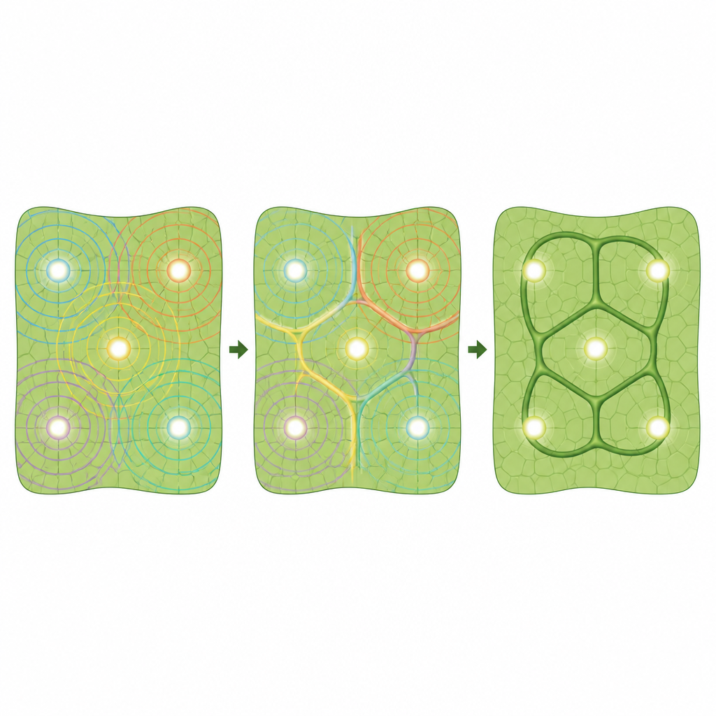
The next question was how living cells could generate such a pattern. Plant biologists have long favored the "canalization" idea, in which the growth hormone auxin flows from sources to sinks and, through feedback with its transport proteins (known as PINs), carves high-flow channels that become veins. Canalization naturally forms tree-like branches that connect sources and drains, but it struggles to explain closed loops that sit between auxin sources instead of linking them. The authors propose a different auxin-based mechanism: hydathodes behave as auxin sources, but instead of forming direct canals, they send out spreading waves of high auxin concentration. Where waves from neighboring hydathodes collide, crest-like ridges appear exactly between them, tracing the eventual paths of major veins.
From model to living leaf
Using computer simulations of a grid of cells, the team showed that when auxin transport is only weakly biased in one direction, waves emerge from each source, move across the tissue, and build ridges at the collision lines. In a two-dimensional leaf-shaped grid seeded with actual hydathode positions, these ridges form loops that closely match both an ideal Voronoi diagram and the real major veins, especially around the leaf edge. The model was refined by adding rules for when vein cells differentiate and how PIN protein levels respond to auxin, which brought simulated PIN patterns into better agreement with microscopy images. Because genetic reporter tools are not yet available in Pilea, the researchers used antibodies that recognize PIN proteins to map where they appear during leaf development. They found strong PIN signal around hydathodes and in primary veins, little signal within secondary veins themselves, and polarized PIN in neighboring cells pointing toward those veins, consistent with the idea of auxin waves shaping the network from hydathode-centered sources.
Why this matters beyond one houseplant
In simple terms, the study concludes that the Chinese money plant draws its looping leaf veins using a geometric rule where each hydathode claims its own "territory," and the borders between territories become major veins. This rule can be produced by chemical waves of auxin that spread from many points and mark their boundaries when they meet. Because similar pores and vein layouts occur in other species, the same wave-and-border mechanism may help explain a wide variety of net-like plant vein patterns. More broadly, the findings show how living tissues can harness straightforward rules of distance and balancing forces to build intricate structures that look complex but are grounded in simple mathematics.
Citation: Zheng, C.X., Palit, S., Venezia, M. et al. Reticulate leaf venation in Pilea peperomioides is a Voronoi diagram. Nat Commun 17, 4111 (2026). https://doi.org/10.1038/s41467-026-71768-3
Keywords: leaf venation, Voronoi pattern, auxin waves, plant geometry, Pilea peperomioides