Clear Sky Science · en
Structures of ZYG11B-EloB-EloC-substrate complex reveal mechanisms of CRL2ZYG11B assembly and function
How Cells Take Out Their Molecular Trash
Every cell in your body constantly builds and destroys proteins. This steady turnover is vital for health, yet the machinery that decides which proteins live or die is incredibly intricate. This study uncovers how one such cellular "bouncer," a protein called ZYG11B, recognizes its targets and works together with partner proteins to mark them for destruction. The findings help explain how cells keep faulty or no‑longer‑needed proteins in check and hint at new ways to harness this system for research and future therapies.
A Cellular Recycling Tag With a Special Signature
Cells use a tag called ubiquitin to label proteins for recycling in a large barrel‑shaped structure that chews them up. Choosing which proteins to tag is the job of E3 ligases, large complexes that act as molecular matchmakers. ZYG11B is one of the parts that gives an E3 ligase its aim. It recognizes proteins that carry a very specific signature at their starting end: a tiny building block called glycine. This so‑called Gly/N degron mark can appear when proteins are trimmed during cell death, when a normal lipid attachment goes wrong, or even in the course of some viral infections, linking ZYG11B to processes such as cell cycle control, immune defense, and the way certain viruses hijack host cells. 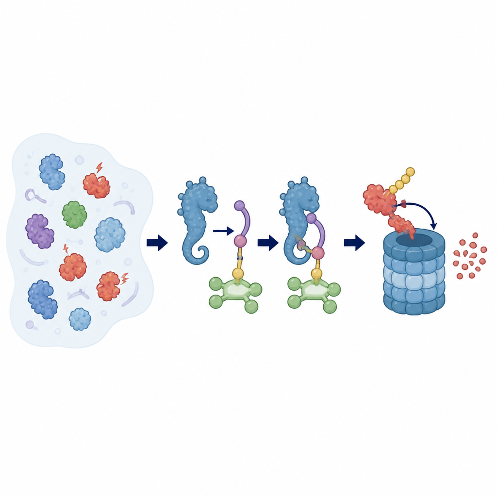
Seeing the Seahorse‑Shaped Worker in Action
To understand how ZYG11B does its job, the researchers used cryo‑electron microscopy, a technique that images frozen molecules in high detail. They solved the structure of full‑length human ZYG11B together with two partner proteins, EloB and EloC, and a short tagged peptide from a viral protein. ZYG11B folds into a striking seahorse‑like shape, with three main regions: a head that docks onto EloB–EloC, a central spine made of repeating units, and a curved tail that cradles the beginning of the target protein. The glycine‑marked peptide nestles into a groove in this tail, with its first three building blocks locked in place through a tight network of contacts, while the rest of the peptide remains more exposed and flexible.
Building the Tagging Machine Piece by Piece
The head of ZYG11B carries a motif that latches firmly onto EloC, which in turn holds EloB, forming a compact adaptor. This adaptor then connects to a larger scaffold protein called Cul2 and a small RING protein that brings in the ubiquitin‑carrying enzyme. ZYG11B uses three separate contact surfaces to grip EloB and EloC, creating a bent loop‑like arrangement that brings the tail’s binding groove close to the catalytic part of the ligase. When the team altered key contact points in ZYG11B’s head or in the groove that holds the glycine‑marked peptide, cells could no longer efficiently destroy proteins bearing that tag. This showed that both substrate binding and adaptor docking are essential for the quality‑control function of this machine.
When One Becomes Two: The Power of Pairing Up
An unexpected twist was that ZYG11B does not always work alone. The structural data revealed that two ZYG11B molecules can pair up back‑to‑back, each gripping its own adaptor and tagged peptide, forming a symmetrical dimer. This pairing involves all three regions of ZYG11B and buries a large contact surface, creating a sturdy assembly with two active binding grooves pointing in opposite directions. The researchers built models of the full ligase in this paired form and showed that it can still accommodate the catalytic components needed for tagging. In test‑tube reactions and cell‑based assays, a mutant version of ZYG11B designed to disrupt dimer formation showed much weaker tagging and degradation of target proteins, indicating that the paired state boosts the efficiency of the system. 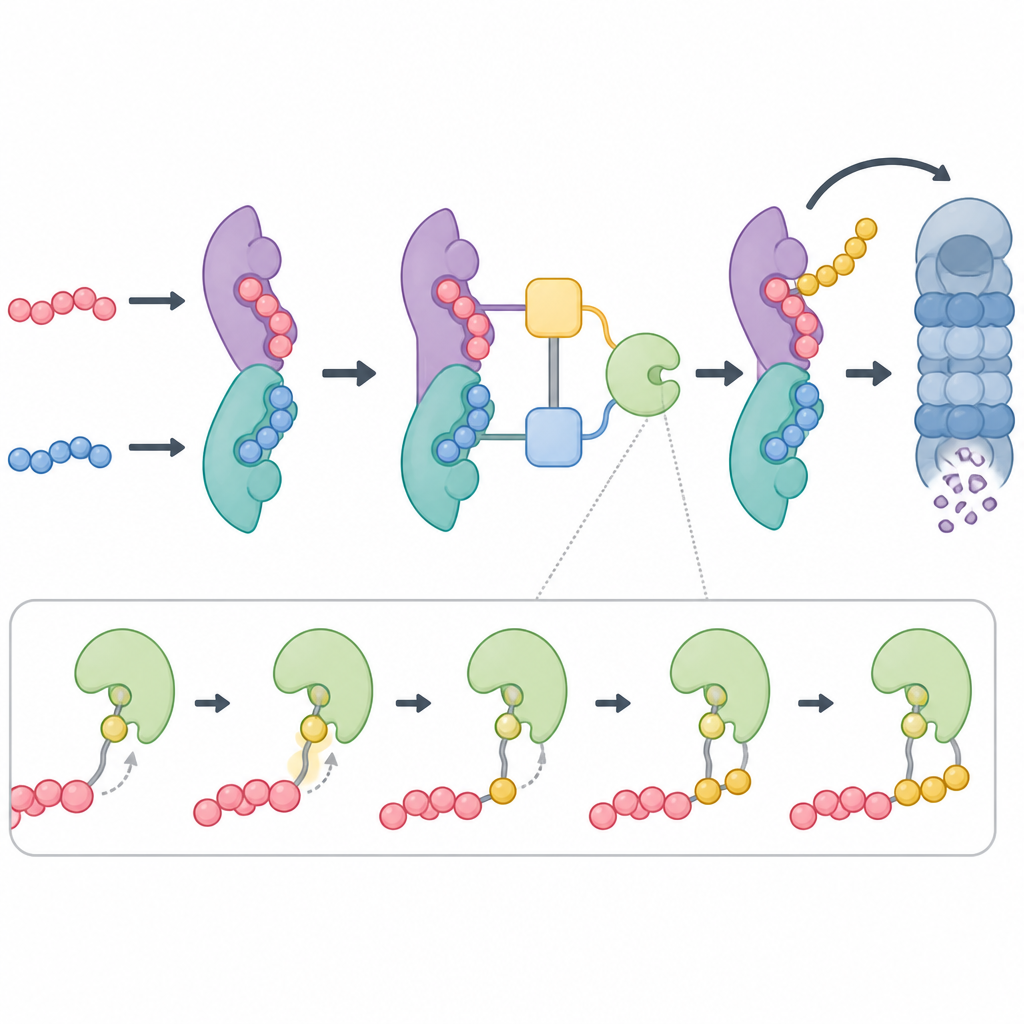
Why These Findings Matter for Health and Design
Together, the results suggest that the ZYG11B‑based ligase can switch between solo and paired states, with both likely present in cells but the paired form playing a leading role in efficient tagging and destruction. By revealing the detailed shape of ZYG11B and exactly how it grips both its partners and its targets, this work provides a blueprint for designing small molecules or custom degraders that recruit ZYG11B to new targets. In the long run, such tools could allow scientists to selectively remove harmful proteins, offering a powerful way to study cellular pathways and potentially address diseases linked to protein quality control.
Citation: Lin, N., Feng, H., Geng, Y. et al. Structures of ZYG11B-EloB-EloC-substrate complex reveal mechanisms of CRL2ZYG11B assembly and function. Nat Commun 17, 4648 (2026). https://doi.org/10.1038/s41467-026-71318-x
Keywords: protein degradation, ubiquitin ligase, ZYG11B, Gly N degron, cryo electron microscopy