Clear Sky Science · en
A controllable human spinal cord model with full dorsoventral patterning
Building a Tiny Human Spinal Cord in the Lab
Understanding how the human spinal cord forms is essential for tackling paralysis, birth defects, and some nerve diseases, but studying these early events directly in embryos is not possible. This research describes a lab-grown, thumb-sized model of the human spinal cord that scientists can precisely control. It mimics how the top and bottom of the spinal cord are shaped by opposing chemical signals, and it even reproduces the behavior of roaming nerve precursors that normally leave the spinal cord to form peripheral nerves.
Why Spinal Cord Patterning Matters
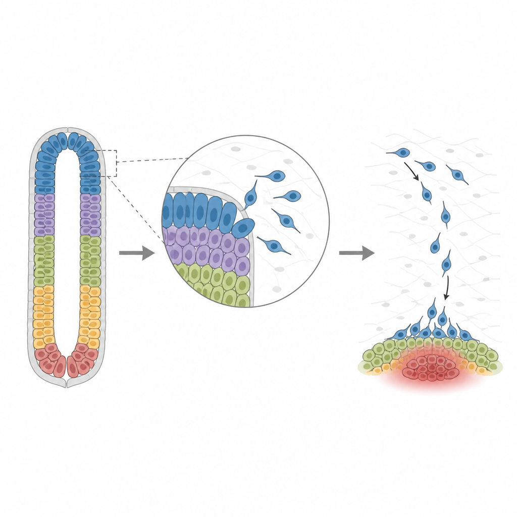
During early development, the spinal cord is organized from back to front into many distinct zones. Each zone eventually produces specific nerve cell types that control movement, sensation, or support functions. At the same time, a special group of cells called neural crest cells bud off from the back side of the forming spinal cord and migrate outward to build structures such as the dorsal root ganglia, which relay touch and pain signals, and the sympathetic ganglia, which help regulate organs. Animal studies have shown that this organization is guided by gradients of signaling molecules, but how this works in humans, and how different signals interact, has been hard to pin down.
A Chip That Engineers Mini Spinal Cords
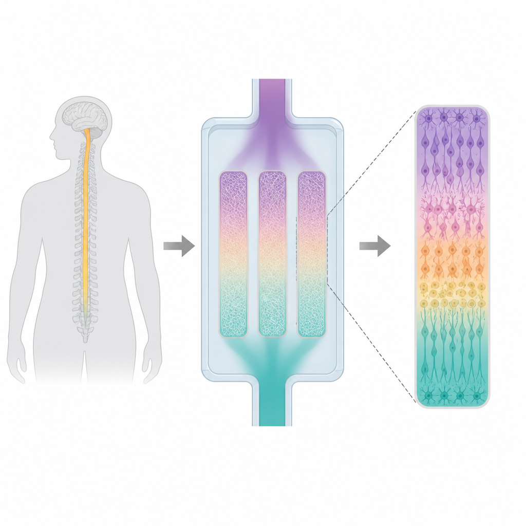
The team created what they call microfluidic spinal cord like structures, or µSCLSs, using human pluripotent stem cells. These stem cells were seeded as neat rectangular colonies on a glass slide and then enclosed inside a narrow channel of soft silicone. The channel was filled with a gel similar to the natural scaffold around the embryonic nervous system. By flowing one set of patterning molecules in from the top of the channel and a different set from the bottom, the researchers produced stable, opposing gradients across each colony. Over twelve days, the cells formed a hollow tube with a central cavity, closely resembling the early neural tube from which the spinal cord arises in embryos.
Recreating the Full Map of the Human Spinal Cord
Careful staining of the µSCLSs showed that they contained all known progenitor zones from back to front, including the most dorsal roof plate, the most ventral floor plate, and the eleven intermediate domains in between. Each zone turned on the same key markers seen in human embryos, and the tissue produced both sensory and motor neurons in the expected locations. Single cell RNA sequencing confirmed that all thirteen major progenitor subtypes present in a human spinal cord at a comparable developmental stage were represented in the model. The cells also displayed human specific gene activity, such as broader expression of certain regulators and early appearance of cells that will become myelin forming support cells, features that differ from mouse development.
Clarifying a Confusing Signal and Watching Cells Migrate
The model allowed the researchers to revisit a long standing puzzle about retinoic acid, a vitamin A derived signal produced by tissues flanking the spinal cord. Previous studies had suggested that retinoic acid could push development toward either dorsal or ventral identities, depending on the setting. By adding or withholding this signal while controlling the other gradients, the team found that retinoic acid generally shifted the pattern toward more dorsal fates, but only when a dorsal signal called BMP was also present. Their analysis pointed to a factor named GDF3 as a key middleman that links retinoic acid to BMP activity and helps tune how ventral motor neuron regions are specified. The µSCLS also faithfully reproduced the behavior of neural crest cells, which emerged from the dorsal side, then some of them migrated downward in a directed way toward a ventral source of a guidance cue called CXCL12; blocking its receptor CXCR4 reduced this ventral migration and shifted cells back toward the dorsal region.
A Versatile Testbed for Future Nerve Research
By showing that a small, engineered tissue can recapitulate full human like spinal cord patterning and the complex movements of neural crest cells, this work provides a powerful testbed for future studies. Researchers can now vary signal strengths, timing, or genes in a controlled manner while observing how specific spinal cord cell types and migrating precursors respond. In the long term, such models could help uncover how certain developmental disorders arise, provide a platform to examine how potential therapies affect human spinal cord cells, and improve our understanding of how a simple sheet of cells self organizes into the intricate wiring that underlies movement and sensation.
Citation: Bok, J., Kim, Y.S., Cheng, F. et al. A controllable human spinal cord model with full dorsoventral patterning. Nat Commun 17, 4539 (2026). https://doi.org/10.1038/s41467-026-71162-z
Keywords: human spinal cord development, stem cell model, morphogen gradients, neural crest migration, retinoic acid signaling