Clear Sky Science · en
A multifaceted model of Entamoeba histolytica KERP2 regulating gene expression and host cell responses
How a Tiny Parasite Can Undermine Our Gut Defenses
Amoebiasis is a gut infection that affects millions of people worldwide, sometimes causing severe diarrhea and life-threatening damage to the intestine. This study looks at how a single parasite protein, called KERP2, can both control the parasite’s own genes and disturb the cells lining our intestines, offering a window into how microscopic invaders outsmart our defenses.
A Close Look at a Gut-Invading Amoeba
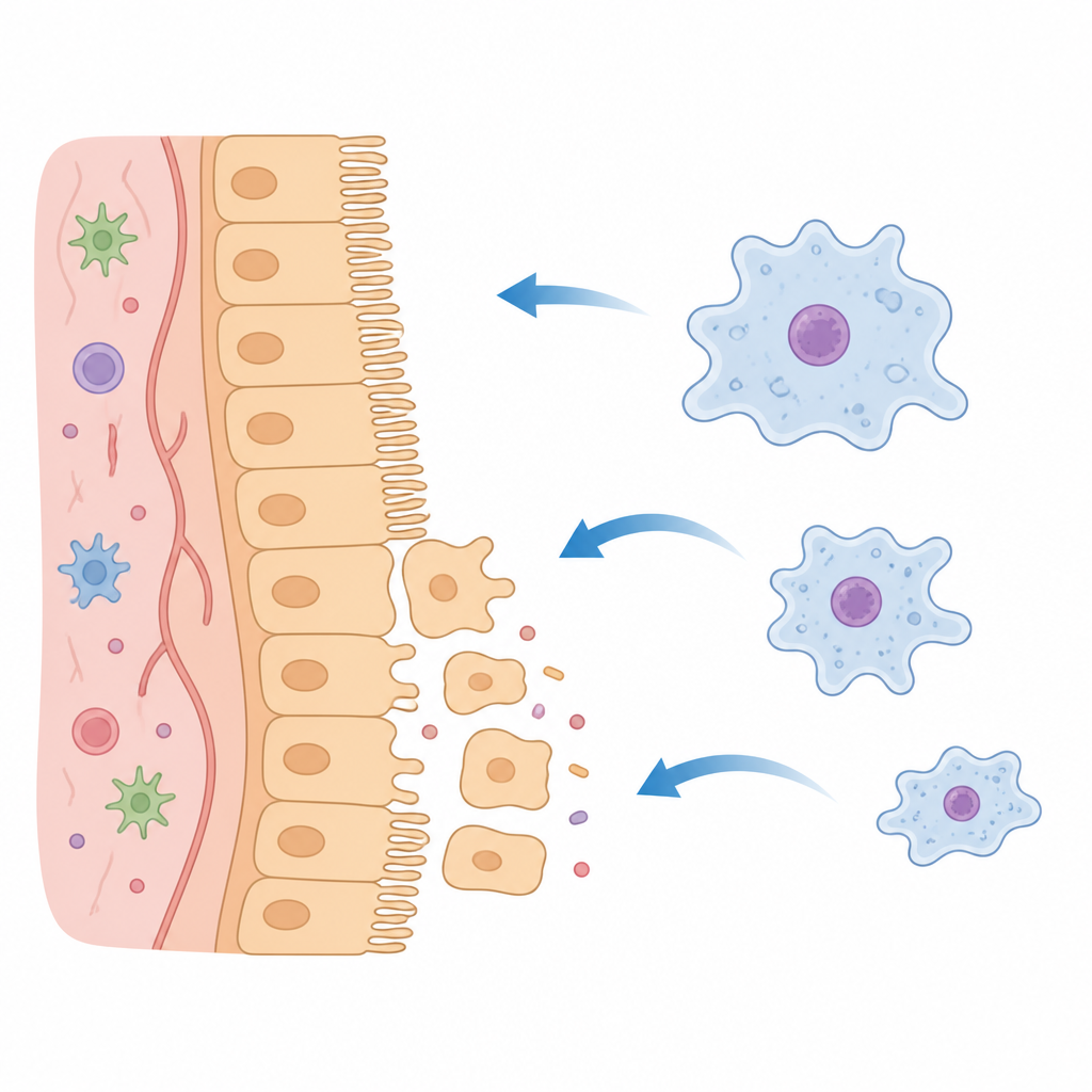
The parasite Entamoeba histolytica lives on the surface of the intestinal wall, where it can inflame and damage the tissue. Unlike microbes that hide inside our cells, this amoeba stays outside but still manages to tamper with host biology. Earlier work had flagged a family of parasite proteins called KERPs as being close to the brush border, the finger-like microvilli on intestinal cells. One of them, KERP2, seemed especially interesting because it is conserved in related amoebae and is linked to how sick people become. Curiously, KERP2 looked like it might be targeted to the parasite’s nucleus, even though it was also recovered from membrane-associated fractions at the cell surface.
A Shape-Shifting Protein Inside the Parasite
Using computer-based sequence comparisons and structural predictions, the authors show that KERP2 has features resembling a chromatin protein called DEK, known in other organisms for shaping how DNA is packed and read. KERP2 contains a SAP-like module that prefers DNA rich in the letters A and T, and a coiled-coil tail that carries a nuclear localization signal. Experiments with tagged versions of KERP2 reveal that the full-length protein accumulates in the parasite nucleus, especially in DNA-dense regions, while a version missing the coiled-coil tail stays mostly in the cytoplasm. In test-tube assays, KERP2 binds strongly to AT-rich DNA but not to GC-rich DNA, and appears to bend or compact the DNA rather than recognizing one exact sequence. Together, these findings paint KERP2 as a chromatin-associated helper that fine-tunes groups of genes, rather than a classic on–off switch.
Tuning the Parasite’s Weapons
To see what KERP2 does for the parasite, the team reduced its production using gene silencing. Parasites lacking KERP2 grew normally, but their gene activity shifted. Many genes tied to amoebic disease, including those for cysteine proteases and pore-forming peptides, were more active, as were genes involved in sulfur and amino acid metabolism. Direct enzyme tests confirmed that cysteine protease activity, a major driver of tissue damage, was higher when KERP2 was knocked down and lower when KERP2 was overproduced. Interaction studies also showed that KERP2 associates with nuclear transport factors, RNA- and DNA-binding proteins, ribosomal components, and trafficking proteins such as Rab11B, hinting that KERP2 sits at the crossroads of gene control and secretory pathways.
Crossing from Parasite to Human Cells
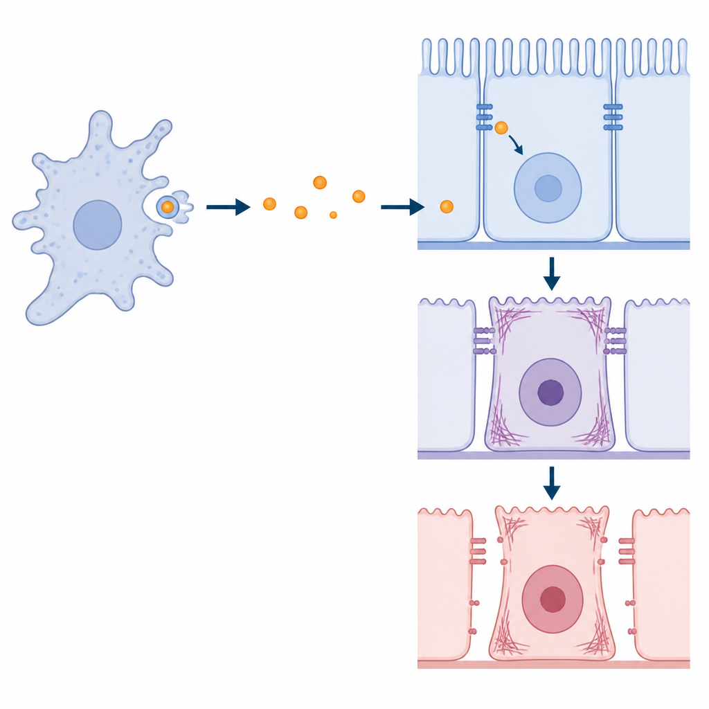
The story does not end inside the parasite. When amoebae contact human intestinal cell lines or a three-dimensional model of human crypts, KERP2 can be detected inside the host cells. Imaging and fractionation show punctate KERP2 signals in host cytoplasm and near microvilli, and small amounts even in the nucleus, although a direct role in host DNA control is not yet proven. Purified KERP2 protein on its own can enter intestinal cells through an energy-dependent process that resembles endocytosis and remains inside for at least two days. Once present in host cells, KERP2 associates with proteins that govern the actin skeleton, cell junctions, and signaling. Host gene profiling reveals shifts in stress responses, metabolism, and pathways linked to cell division and structural organization.
Rewiring Cell Shape and Barrier Strength
Functionally, KERP2 alters how intestinal cells behave and how well they hold together. Cells exposed to KERP2, either via live parasites or added purified protein, show increased DNA synthesis, suggesting a nudge toward cell-cycle activity. Their actin fibers reorganize, cells become more elongated, and the usual dense actin ring at cell edges weakens. In wound-healing assays designed to track how fast a sheet of cells closes a gap, KERP2 slows collective movement when new cell division is limited. Measurements of electrical resistance across cell layers and tracking of fluorescent molecules crossing the barrier reveal that KERP2 can reduce ion-tight sealing and, in some cases, increase leakage of larger molecules through the junctions. Interestingly, when the parasite lacks KERP2, it compensates with higher protease activity, causing major drops in electrical resistance but not always in large-molecule leak, implying the host cytoskeleton can sometimes tighten up to counteract the damage.
What This Means for Understanding Infection
This work suggests that KERP2 is a dual-purpose tool: inside Entamoeba, it helps keep virulence genes and metabolism in balance, while during contact with the gut lining it can be transferred into host cells to tweak their structure, growth, and barrier properties. Rather than acting purely as a toxin, KERP2 seems to fine-tune how rigid or leaky the intestinal surface is, potentially helping the parasite adapt to different host environments. Although more research is needed, especially in animal models, the study offers a broader view of how extracellular parasites can use multi-tasking proteins to coordinate their own gene programs with subtle remodeling of host tissues.
Citation: Peng, R., Santos, H.J. & Nozaki, T. A multifaceted model of Entamoeba histolytica KERP2 regulating gene expression and host cell responses. Nat Commun 17, 4433 (2026). https://doi.org/10.1038/s41467-026-70847-9
Keywords: Entamoeba histolytica, amoebiasis, intestinal epithelium, host pathogen interaction, parasite virulence