Clear Sky Science · en
High-fidelity single-frame computational super-resolution using signal-preserving denoising-enabled deconvolution
Sharper Views Inside Living Cells
Modern microscopes have transformed biology, but many of life’s most important structures are still too small or too faint to see clearly. This paper introduces a new way to turn ordinary fluorescence microscopes into powerful nanoscale imaging tools using only software. By cleaning up noisy pictures in a very careful way and then mathematically “unblurring” them, the method reveals details such as fine actin fibers, mitochondrial membranes, and nuclear pores in single snapshots of living cells—without special dyes or exotic optics.
Why Small Structures Are Hard to See
Inside cells, proteins assemble into filaments, rings, and tiny clusters that control movement, communication, and disease. Yet conventional light microscopes are limited by diffraction, which blurs details smaller than a couple of hundred nanometers. Optical super‑resolution systems can beat this limit, but they usually need complex, expensive hardware, intense light that can damage cells, and long recording times that miss fast events. Software‑based super‑resolution promises to upgrade existing microscopes instead, but current approaches have trade‑offs: traditional deconvolution algorithms amplify noise and create false features, while deep‑learning methods often work only on structures similar to those they were trained on.
A Two-Step Path From Blur to Detail
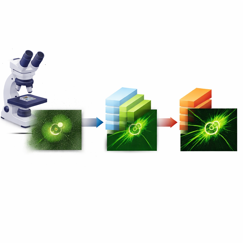
The authors propose a simple but powerful two‑step strategy called 3Snet‑CLID. First, a deep‑learning network is used purely as a denoiser: it takes a single noisy image and outputs a much cleaner version, with random speckles and background greatly reduced. Second, a well‑known mathematical procedure called Richardson–Lucy deconvolution is applied to this cleaned image to undo the microscope’s blur and produce a super‑resolved result. Crucially, the denoising network is designed to preserve the exact distribution of brightness at each pixel instead of smoothing it across neighboring pixels. This careful conservation of signal statistics means that the later deconvolution step can push resolution much further without generating obvious artifacts.
Listening to Each Pixel on Its Own
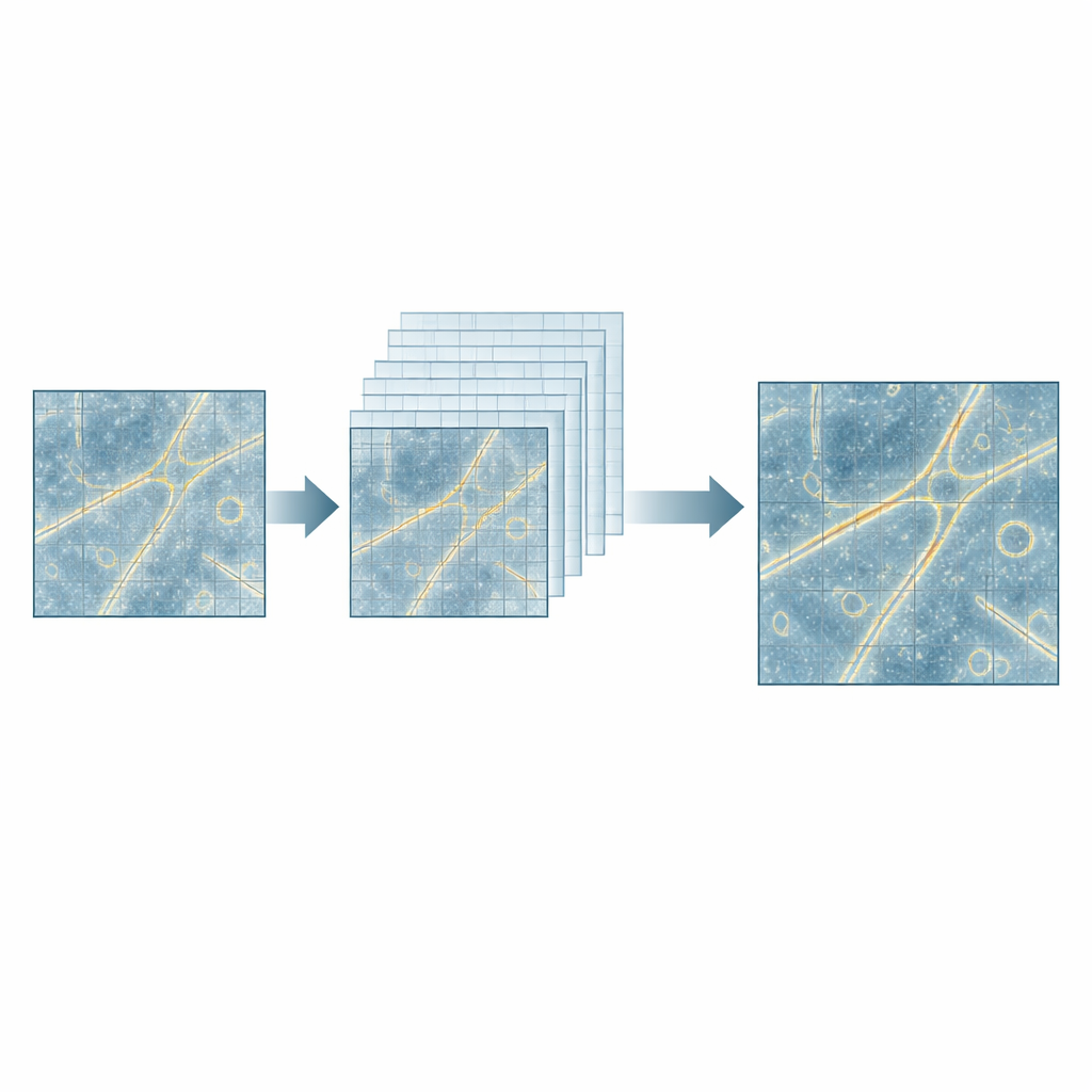
To train such a faithful denoiser, the team first needed nearly noise‑free reference images that still reflected the true intensity at every pixel. They achieved this with a clever per‑pixel strategy they call single‑pixel‑synchronized switching, or 3S. Using special fluorescent proteins that can be turned ON and OFF with light, they collect many frames in each state. By averaging repeated ON images, they reduce random noise; by subtracting the averaged OFF image from the averaged ON image, they remove fixed background patterns. Because each pixel is processed independently, the underlying brightness pattern is preserved. These “clear” images serve as ground truth to train a U‑net‑style deep‑learning model that combines supervised learning (using the clear images) and self‑supervised learning (using pairs of noisy images), yielding a robust, structure‑agnostic denoiser called 3Snet.
Proving the Method on Test Patterns and Real Cells
The researchers rigorously tested 3Snet‑CLID on both synthetic and experimental samples. On simulated microtubule patterns and commercial line grids, the method cleanly separated features spaced as close as 60–65 nanometers—far below the normal diffraction limit and well beyond what standard widefield images, popular denoising networks, or even advanced sparse deconvolution could resolve. Fluorescent beads of 20–100 nanometers in size provided a second, independent check of resolution. In biological specimens, 3Snet‑CLID turned noisy widefield or spinning‑disk confocal images of actin networks, endoplasmic reticulum, and mitochondria into crisp views with roughly fivefold resolution gains. It resolved ring‑shaped nuclear pores whose sizes matched electron‑microscopy standards and uncovered dynamic events such as the remodeling of mitochondrial outer membranes and the interplay between actin flows and microtubule growth during immune synapse formation in T cells.
A Software Upgrade for Everyday Microscopes
From a practical standpoint, the advance lies in turning single, quickly acquired frames into high‑fidelity nanoscale images, using common fluorescent labels and standard microscopes. Because the network focuses on denoising while preserving the true brightness pattern, and leaves the sharpening to a physics‑based deconvolution step, it generalizes well across many structures without heavy parameter tuning. Under typical conditions, the approach achieves around 60‑nanometer resolution with minimal artifacts, enabling researchers to watch fine cellular structures evolve in real time. For non‑specialists, this work shows that smarter image processing alone can unlock a wealth of hidden detail in familiar microscope pictures, bringing ultrasmall cellular features into everyday reach.
Citation: Xue, F., Yuan, L., He, W. et al. High-fidelity single-frame computational super-resolution using signal-preserving denoising-enabled deconvolution. Nat Commun 17, 4056 (2026). https://doi.org/10.1038/s41467-026-70791-8
Keywords: super-resolution microscopy, image denoising, deep learning imaging, live-cell imaging, fluorescence microscopy