Clear Sky Science · en
Structural basis of human γTuRC closure during CM1-activated microtubule nucleation
How cells build their inner scaffolding
Inside every animal cell, a hidden scaffold made of tiny tubes helps move chromosomes during cell division, ship cargo, and shape the cell itself. This study reveals how a key cellular machine, which starts these tubes, flips from an idle to an active state. Understanding this microscopic switch offers insight into how cells keep their internal architecture reliable and on time.
The starting platform for tiny tubes
Microtubules are hollow tubes built from repeating protein building blocks that assemble into 13 side-by-side strands. Getting this process started is hard, so cells rely on a ring-shaped platform called the gamma-tubulin ring complex, or gammaTuRC. This large protein assembly acts like a template that helps the first row of building blocks line up. In simple organisms such as yeast, gammaTuRC is put together directly where it is needed, at a cellular structure that organizes the microtubule network. In human cells, however, gammaTuRC is pre-built in the fluid interior of the cell and then shipped to different organizing centers. To avoid stray tube formation, the human version is held in a bent, open shape that does not match the geometry of a proper microtubule and therefore remains mostly inactive.
A helper that steps on the gas
Several cellular proteins can boost the activity of gammaTuRC. Many of them share a short region called CM1, which binds directly to the ring complex. Using a sensitive microscope that records individual growth events, the authors watched purified human gammaTuRC molecules on a glass surface as they tried to launch microtubules. On their own, the complexes were sluggish. When the CM1 fragment from a human protein was added, nucleation sped up dramatically, by more than a hundredfold with normal tubulin building blocks and even more when a specially designed tubulin variant that favors growth was used. At high CM1 levels, nearly every gammaTuRC on the surface eventually fired, showing that this helper can fully activate the entire population.
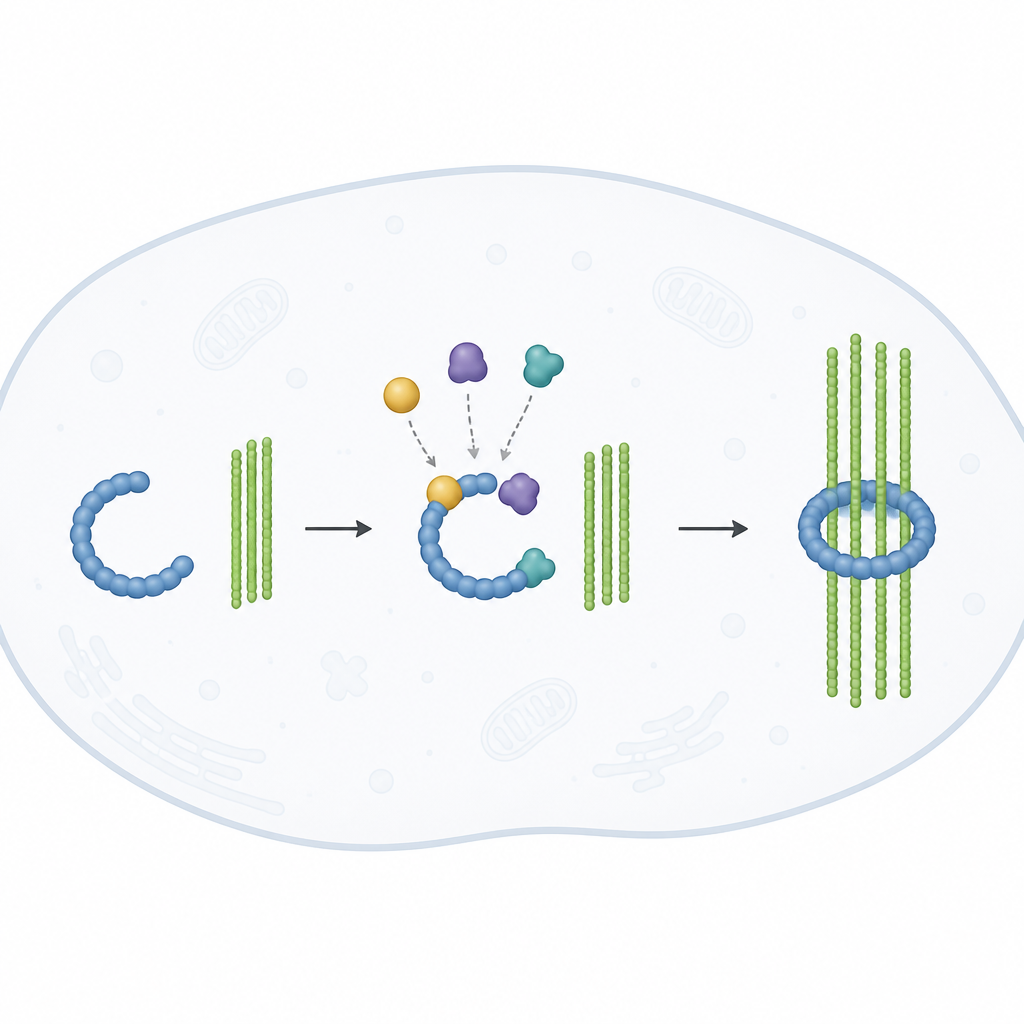
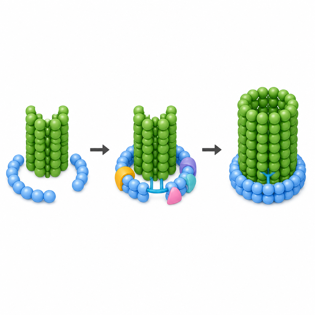
Synergy between binding and tube growth
By tagging CM1 with a fluorescent marker, the researchers could time when it attached to individual gammaTuRC molecules and when each microtubule began to grow. Sometimes the tube appeared as soon as CM1 bound, but often there was a delay of several minutes. This suggested that CM1 binding alone was not enough: the complex also had to shift through different shapes before a new tube could take off. The team proposed that CM1 primes gammaTuRC, making it easier for the first row of tubulin building blocks to assemble. The act of tube growth then helps drive the complex into a fully closed, symmetric ring that matches the 13-strand structure of a normal microtubule. In other words, the template and the growing tube cooperate to complete the switch from off to on.
Snapshots of the ring snapping shut
To see these shape changes in detail, the authors turned to cryo-electron microscopy, a method that images flash-frozen molecules at near-atomic resolution. They captured gammaTuRC bound to CM1 while it was already capped by the base of a freshly formed microtubule, using either normal tubulin or the growth-friendly mutant. In both cases, the ring complex was completely closed and its components lined up in a regular pattern that matched a standard 13-strand tube. This confirmed that, at least in human cells, efficient nucleation involves full closure of the ring. Earlier studies in frogs had hinted that vertebrate gammaTuRC might stay partly open, leading to unusual tube shapes, but the new work argues that human complexes do achieve a perfect fit when actively nucleating.
The latch and inner brace that lock the ring
At higher resolution, the authors could identify specific protein segments that act like hardware to lock the ring shut. A flexible extension from one gammaTuRC subunit, working together with a small partner protein, forms a structure they call the latch. This latch reaches from the trailing end of the open spiral to the opposite side, gripping both the first gamma-tubulin in the ring and the first alpha-tubulin in the emerging microtubule. In parallel, CM1 dimers form bridges between neighboring subunits around the outside of the cone, with especially strong contacts at one special site. From there, an extra loop stretches across the seam to touch gamma-tubulin on the far side. Inside the cone, an actin molecule, part of an internal brace, swings into a new position so it no longer blocks closure and instead contacts the terminal subunit, helping stabilize the shut ring.
Why this molecular switch matters
To a non-specialist, the message of this work is that human cells use an elegant safety mechanism to control when and where they build their internal tubes. The gammaTuRC machine is assembled in a safe, inactive shape. A helper region called CM1 docks onto it and loosens it up, but full activation only happens when the first tubulin building blocks arrive and a microscopic latch and brace lock the ring into a perfect circle. This combined action ensures that new microtubules start with the right geometry and at the right locations, supporting accurate cell division and orderly organization inside our cells.
Citation: Serna, M., Brito, C., Speroni, S. et al. Structural basis of human γTuRC closure during CM1-activated microtubule nucleation. Nat Commun 17, 4488 (2026). https://doi.org/10.1038/s41467-026-70773-w
Keywords: microtubule nucleation, gammaTuRC, CM1 motif, cryo electron microscopy, cell division