Clear Sky Science · en
Ceramide disrupts TM9SF2-PGK1 axis to redirect PD-L1 trafficking and enhance antitumor immunity
Why this research matters for cancer treatment
Many successful cancer drugs work by lifting the brakes that tumors place on the immune system, yet not all patients benefit and some tumors regain the upper hand. This study uncovers a hidden cellular "sorting" system that controls how much of a key immune brake, called PD-L1, sits on the surface of cancer cells. By revealing how a natural fat molecule can flip this sorting switch, the work points to new ways to help immune therapies work better and for longer.
The immune brake on tumor-fighting cells
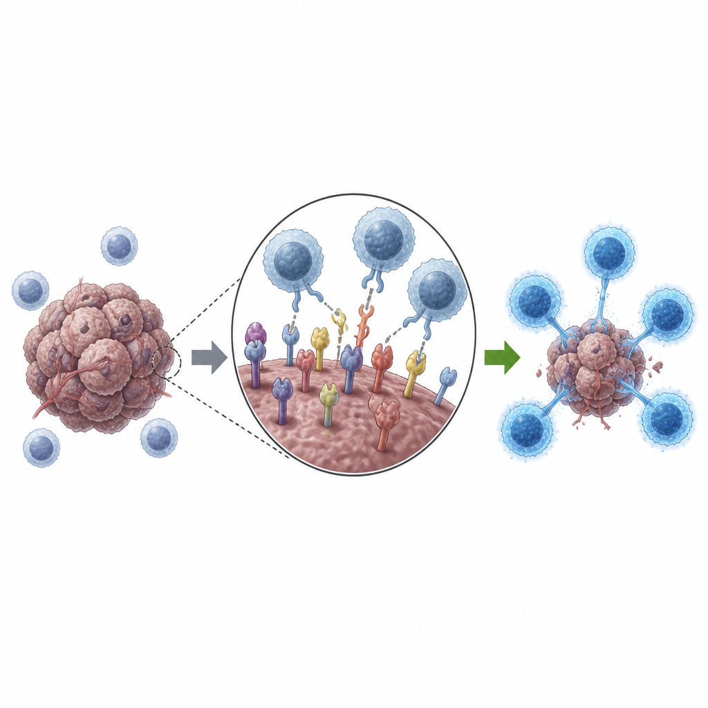
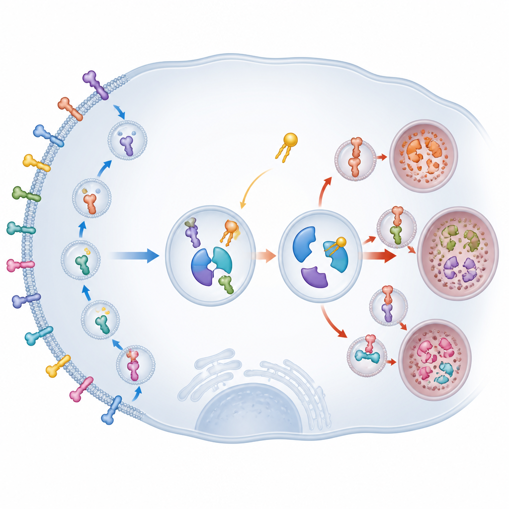
Our immune system relies on killer T cells to recognize and destroy cancer cells, but tumors often defend themselves by displaying PD-L1 on their surface. When PD-L1 engages the PD-1 receptor on T cells, it dampens their attack. Existing checkpoint drugs block this interaction from the outside. However, cancer cells constantly pull PD-L1 in from the surface and send it back out again, or send it to cellular "recycling" or "trash" compartments. This constant shuttling can weaken the effect of antibody drugs and contribute to resistance, so scientists are keen to understand and control how PD-L1 is routed inside the cell.
A trafficking hub that keeps PD-L1 on the surface
Using a CRISPR gene-editing screen focused on fats called sphingolipids, the researchers identified a little-studied protein, TM9SF2, as a crucial keeper of PD-L1 levels. When TM9SF2 or its related family members were removed from lung cancer cells, PD-L1 on the surface and inside the cells dropped sharply, even though the gene that encodes PD-L1 was unchanged. These TM9SF proteins instead slowed the breakdown of PD-L1, allowing it to persist longer. In both cell cultures and mouse tumors, loss of TM9SF2 made cancer cells far more vulnerable to attack by CD8 T cells, boosting their ability to kill tumor cells and produce alarm signals such as interferon-gamma.
How an internal conveyor system is rewired
The team then discovered that TM9SF2 forms a complex with another protein, PGK1, better known for its role in energy production. Inside tumor cells, TM9SF2 and PGK1 join with a recycling factor called RAB11 at endosomes, the sorting stations of the cell. Together, they act like a conveyor system that routes internalized PD-L1 back to the cell surface instead of to waste compartments. When TM9SF2 or PGK1 was disabled, PD-L1 was diverted into lysosomes, the cell’s digestive sacs, where it accumulated and was broken down. At the same time, a separate protein, HIP1R, which normally ferries PD-L1 to lysosomes, became more stable because PGK1 was no longer tagging it for destruction. In essence, the TM9SF2–PGK1 pair both promotes recycling of PD-L1 and dismantles the machinery that would otherwise send it to the trash.
A natural fat that breaks the complex and frees T cells
Lipid profiling showed that lowering TM9SF2 levels altered several ceramides, a family of waxy fat molecules. Among them, one species, Cer(d18:1/26:0), stood out. When this ceramide was added to tumor cells, PD-L1 levels on the surface and inside the cells fell, while HIP1R became more stable. Further experiments revealed that this ceramide weakens the binding between TM9SF2 and PGK1, breaking apart the recycling hub. As a result, PD-L1 is steered away from recycling endosomes toward lysosomes and degraded. In mouse models of melanoma and leukemia, treatment with this ceramide shrank tumors, lowered PD-L1 on cancer cells, and increased the number and activity of tumor-infiltrating CD8 T cells.
Implications for patients with lung and other cancers
When the authors examined patient data, TM9SF2 and PGK1 were frequently elevated in lung adenocarcinoma, and higher TM9SF2 levels were linked to poorer survival in leukemia. Tumor samples with more TM9SF2 also tended to have more PD-L1. Importantly, cancer cells freshly isolated from lung tumors responded to a PGK1-blocking drug or to the ceramide by lowering PD-L1 and redirecting it to lysosomes. Together, these findings suggest that targeting the TM9SF2–PGK1 complex, or using specific ceramides to destabilize it, could complement current checkpoint inhibitors. For patients, this strategy aims not to invent a new immune brake, but to quietly remove the spare parts that tumors rely on to keep that brake working.
Citation: Zheng, Y., Yang, F., Wang, M. et al. Ceramide disrupts TM9SF2-PGK1 axis to redirect PD-L1 trafficking and enhance antitumor immunity. Nat Commun 17, 4525 (2026). https://doi.org/10.1038/s41467-026-70764-x
Keywords: PD-L1 trafficking, cancer immunotherapy, ceramide, TM9SF2 PGK1, CD8 T cells