Clear Sky Science · en
Anterior and posterior retrosplenial cortex form distinct visuospatial circuits in the mouse
How the brain keeps us oriented
Finding your way around a room or a city may feel effortless, but it depends on brain circuits that constantly blend what you see with how you move. This study looks at a little-known brain region in mice, the retrosplenial cortex, and shows that its front and back parts play different roles in turning sights and sensations into a sense of place.
Two sides of the brain’s inner compass
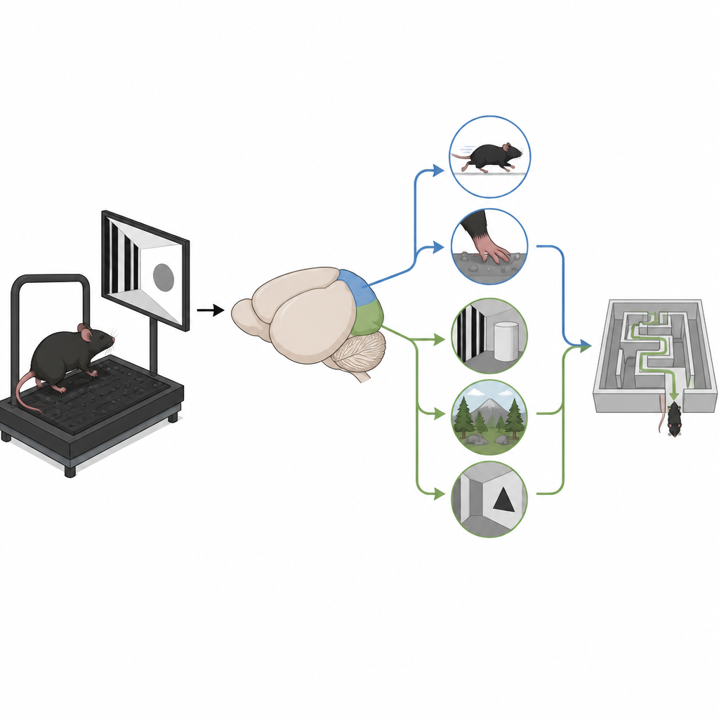
The retrosplenial cortex sits near the back of the brain and helps connect memory, vision, and movement. The researchers asked whether its front (anterior) and back (posterior) halves handle space in the same way. Using tiny microscopes to watch thousands of nerve cells in awake mice running on a treadmill, they tracked how activity changed as the animals moved along a track marked with touch cues and visual scenes. They also traced long-distance wiring across the whole brain to see where inputs to each half came from. Together, these tools let them relate what each area does to the information it receives.
Front section: sharper sense of position
When mice ran along a belt with a fixed reward spot and touch landmarks, many cells in the front retrosplenial cortex lit up at specific locations along the track. These responses were sharp and reliable, allowing the researchers to read out the mouse’s position with only a few centimeters of error. Removing the touch landmarks reduced this precise coding mainly in the front section, showing that it leans more on tactile information. Even in the dark, when visual cues were gone, front cells still carried clearer position signals than those in the back section, suggesting a strong link to movement and body-based cues.
Back section: vision-rich maps of space
The back retrosplenial cortex told a different story. In the simple touch-marked setup, its position signals were weaker and more spread out along the track. But when the mice moved through a visually rich virtual corridor filled with clear landmarks, cells in the back section showed much stronger tuning to position, rivaling the front section. The same area also contained more cells that responded reliably to moving patterns on a screen, and these cells preferred slow, fine visual details, like narrow bars drifting slowly. In contrast, visual cells in the front section were more sensitive to fast, coarse motion, suggesting that each side emphasizes different kinds of visual information.
Distinct wiring for touch, vision, and memory
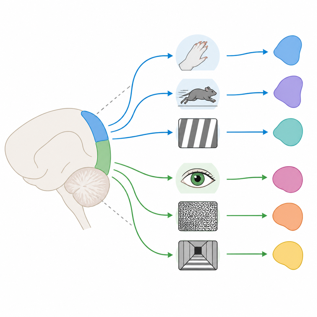
To understand why these differences arise, the team injected tracers into the front and back retrosplenial cortex and mapped all the brain regions that sent inputs. The front half received more connections from motor and touch areas, which track running and contact with the environment, as well as parts of the hippocampal memory system linked to precise spatial layouts. The back half drew stronger input from primary and posteromedial visual areas that process detailed scenes, along with different memory and thalamic regions tied to context and emotion. This wiring pattern mirrors the functional split: the front section integrates body motion and touch with space, while the back section is more tightly coupled to vision and scene context.
Why this matters for understanding navigation
Taken together, the results reveal a front-to-back gradient inside a single brain region that helps animals know where they are. The front retrosplenial cortex behaves like a hub for precise, movement- and touch-based estimates of position, while the back portion specializes in using rich visual scenes to anchor those estimates. By showing how these complementary circuits are organized and connected, the study offers a clearer picture of how the brain combines different senses to build a stable inner map of the world.
Citation: Wei, YT., Couto, J., Kloosterman, F. et al. Anterior and posterior retrosplenial cortex form distinct visuospatial circuits in the mouse. Nat Commun 17, 4388 (2026). https://doi.org/10.1038/s41467-026-70762-z
Keywords: spatial navigation, retrosplenial cortex, visual landmarks, mouse brain circuits, position coding