Clear Sky Science · en
Suppression of pathological oscillations with transcranial focused ultrasound in Parkinson’s disease
Why this matters for people with Parkinson’s
People with Parkinson’s disease often cope with slowed movement, stiffness and tremor that current drugs and brain implants can only partly relieve. This study explores whether gentle pulses of focused ultrasound delivered from outside the skull can quiet abnormal brain rhythms linked to these symptoms, pointing toward a future treatment that does not require brain surgery. 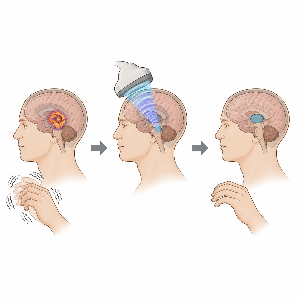
A new way to reach deep brain areas
Parkinson’s disease alters how groups of brain cells fire together, especially in deep structures that help control movement. In these areas, a type of brain rhythm called beta activity becomes unusually strong and is tied to slower movement. Doctors can already ease symptoms by replacing dopamine with medication or by using deep brain stimulation, which involves surgically implanted electrodes that deliver electrical pulses. However, surgery is invasive and not suitable for everyone. Focused ultrasound offers a different route: sound waves are steered through the skull to precise points inside the brain without any incision.
Designing a careful proof of concept
The researchers worked with four men who already had deep brain stimulation electrodes in a region called the subthalamic nucleus. This allowed the team to record deep brain signals while testing ultrasound. Using brain scans and advanced mapping methods, they identified a connected region in a nearby structure called the globus pallidus as the ultrasound target. Each participant took part in two sessions on separate days. On one day, ultrasound pulses were focused on this movement control hub. On the other, pulses were directed toward a fluid-filled space in the brain used as an active control site. Within each day, sessions with no ultrasound were followed by sessions with ultrasound, and both the volunteers and the examiners were kept unaware of which condition was active.
Quieting harmful rhythms and speeding reactions
When ultrasound was aimed at the movement control region and pulsed at 130 times per second, the abnormal beta rhythm recorded from the nearby deep brain electrode on the same side fell on average by about ten percent compared with the control condition. In volunteers who showed two distinct beta peaks, only the lower-frequency part was reduced, matching what is known about which pathways contribute most to movement slowing. The drop in deep beta activity closely tracked changes in activity measured over the surface of the brain in the primary motor area on the same side, suggesting that ultrasound influenced a wider movement network. Crucially, during a decision and reaction task in which participants judged the motion of dots on a screen, their reaction times improved by nearly eighteen percent when ultrasound was applied to the movement hub.
Ruling out simple artefacts
Because ultrasound can cause subtle mechanical vibrations, the team carefully checked whether the observed signal changes were real brain effects rather than technical noise. In bench experiments, they sonicated a deep brain electrode placed in a gel model and tested different pulse patterns. Slow pulse rates created clear mechanical artefacts in the frequency range of interest, but the fast pattern used in the main study did not. Simulations also showed that any heating in the brain tissue stayed well within accepted safety limits, and the ultrasound focus was kept several millimetres away from the implanted electrode tips. These steps support the conclusion that the reductions in beta power and faster reactions reflect genuine changes in brain activity. 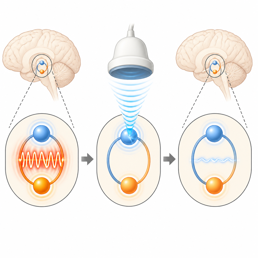
What this could mean for future treatment
This small, early study suggests that focused ultrasound, tuned to match successful electrical stimulation, can damp down harmful brain rhythms tied to movement slowing in Parkinson’s disease and modestly improve performance on a movement task, all without surgery. While the work involved only four people and did not show clear changes on standard bedside rating scales, it offers a first demonstration that noninvasive ultrasound can push a well-established disease signal in the same helpful direction as existing therapies. With further research to refine the pulse settings, understand why responses vary between individuals, and test longer-lasting effects, this approach could become part of a new toolkit for treating Parkinson’s and perhaps other brain disorders driven by abnormal rhythms.
Citation: Eraifej, J., Toth, J., Hanemaaijer, J. et al. Suppression of pathological oscillations with transcranial focused ultrasound in Parkinson’s disease. Nat Commun 17, 4471 (2026). https://doi.org/10.1038/s41467-026-70714-7
Keywords: Parkinson’s disease, focused ultrasound, deep brain stimulation, brain rhythms, neuromodulation