Clear Sky Science · en
Ultra-precision deconvolution of spatial transcriptomics decodes immune heterogeneity and fate-defining programs in tissues
Seeing Cells in Their Neighborhoods
Our organs are built from many kinds of cells packed tightly together, and their exact positions matter for how diseases like cancer grow or how wounds heal. This study introduces a new way to read gene activity maps of tissues so precisely that scientists can pinpoint small, rare immune cells and understand how they team up with nearby cells to fight tumors or form scars.
Sharper Maps from Blurry Signals
Modern spatial gene mapping tools measure which genes are active at thousands of tiny spots across a tissue slice. The catch is that each spot usually contains several cells mixed together, so the signal is blurry. The researchers developed UCASpatial, a computer method that teases apart this mixture and estimates how many cells of each type are present at every spot. It learns typical gene patterns from single-cell datasets, then gives extra weight to genes that are especially good at distinguishing one cell type from another, using ideas from information theory to decide which genes carry the clearest identity clues.
Testing the Method in Virtual Tissues
Before trusting UCASpatial on real samples, the team built simulated tissues where the true cell makeup was known. They mixed gene profiles from different immune, tumor, and support cells into artificial spots of varying complexity and density. Across many tests, UCASpatial consistently made more accurate guesses about cell proportions than several leading methods, particularly when cell types were very similar to one another or present at very low levels. It remained reliable even when spots contained many cells or when there were many closely related immune subgroups. 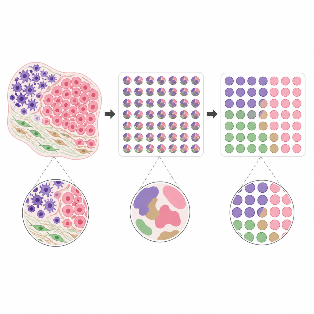
Immune Deserts in Colon Cancer
The researchers then applied UCASpatial to human colorectal cancer samples. By combining detailed maps of immune cells with inferred genetic changes in tumor cells, they tracked how different cancer clones shaped their surroundings. Some clones sat in regions rich in T cells, while others created T cell–poor “immune deserts.” A recurring feature of these T cell–excluded zones was extra copies of a stretch of chromosome 20 called 20q. Tumors with this gain tended to shut down a particular family of ancient viral-like DNA elements known as HERV-H and showed weaker antiviral and alarm signals that normally help draw killer T cells into tumors. Patients whose tumors carried this 20q change also fared worse on immune checkpoint therapy, hinting that this genetic shift helps tumors hide from the immune system.
Cell Communities that Heal or Scar
UCASpatial also decoded how wounds heal in the ears of two mouse strains: one that perfectly regenerates tissue and one that heals with a scar. By following many cell types over time and space, the team saw that early influxes of white blood cells were similar in both strains, but later patterns diverged. In the scarring strain, a tight-knit trio formed inside the wound bed: a special cartilage-like cell (Igfbp5+ chondrocyte), a scar-associated fibroblast, and a lipid-handling macrophage. Together, these three cell types built up dense connective tissue. Signals carried by the molecule IL-11 and its receptor were especially strong in this triad. When the researchers blocked IL-11’s receptor or nudged fibroblasts away from a scar-forming state, this pro-scar community shrank and the wounds closed more cleanly, with better regeneration. 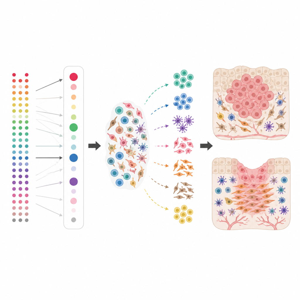
What These Findings Mean
By turning fuzzy gene maps into sharp cellular blueprints, UCASpatial lets scientists link specific genetic changes and cell neighborhoods to real outcomes, such as T cell–poor tumors or scarred skin. For a general reader, the key message is that where cells sit, who their neighbors are, and which signals they exchange can decide whether a tumor is invisible to the immune system or whether a wound regenerates or scars. Tools like UCASpatial help uncover these hidden patterns, opening the door to more targeted cancer therapies and smarter strategies to promote true tissue repair.
Citation: Xu, Y., Huang, Z., Zhang, Y. et al. Ultra-precision deconvolution of spatial transcriptomics decodes immune heterogeneity and fate-defining programs in tissues. Nat Commun 17, 4269 (2026). https://doi.org/10.1038/s41467-026-70645-3
Keywords: spatial transcriptomics, immune microenvironment, colorectal cancer, wound healing, single-cell analysis