Clear Sky Science · en
Microglial cathepsin B is necessary for neuronal efferocytosis in zebrafish and mice during brain development
Why Brain Cell Cleanup Matters
As the brain develops, it produces far more nerve cells than it ultimately needs. Roughly half of these newborn neurons are quietly removed, a process that must be both efficient and gentle so that healthy circuits emerge. This study asks a deceptively simple question with big implications: how do the brain’s resident immune cells dispose of all these dying neurons without getting overwhelmed, and what happens when this cleanup machinery fails?
The Brain’s Janitors at Work
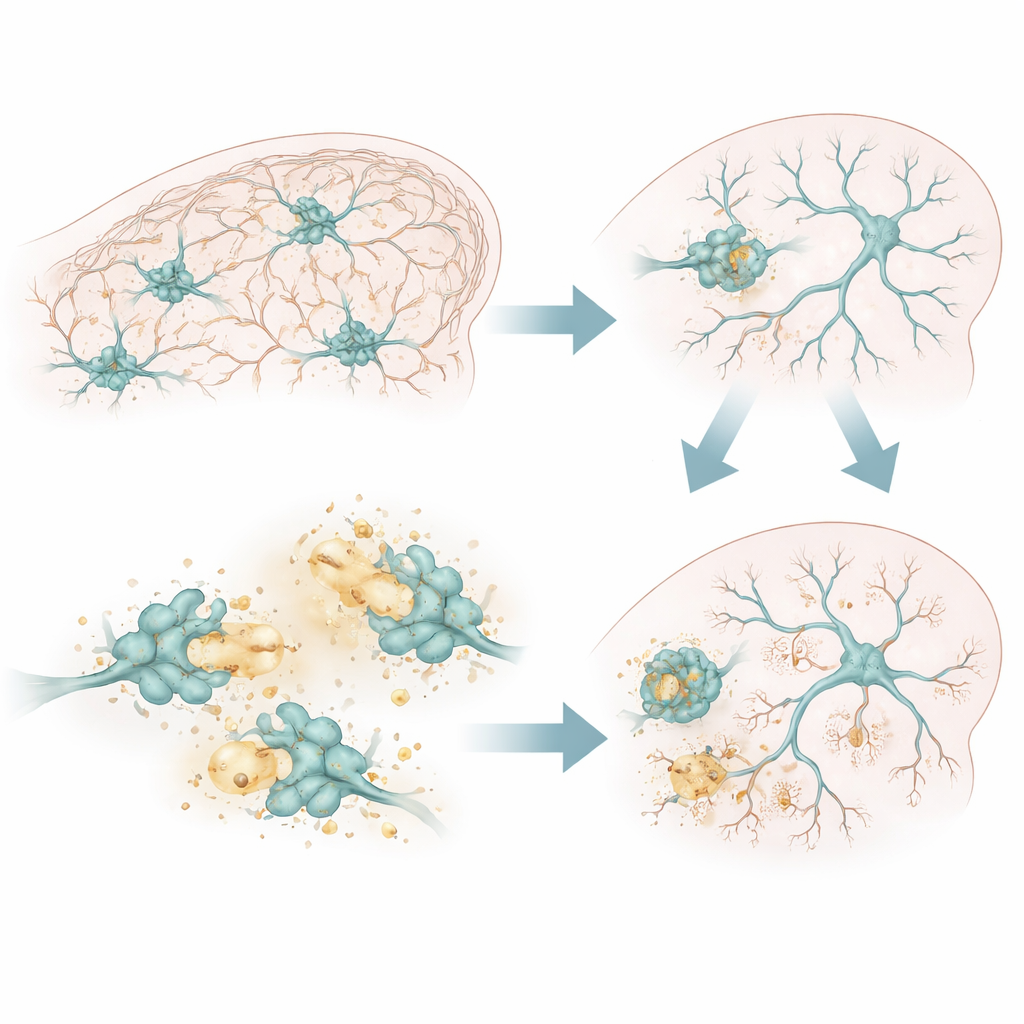
Specialized immune cells called microglia patrol the brain, constantly extending and retracting their branches to sense their surroundings. When a neuron reaches the end of its life, it displays surface signals that act like a “pick me up” flag for microglia. The microglia then engulf the dying cell whole in a process known as efferocytosis, pulling it into an internal bubble that later fuses with acidic recycling compartments. Inside these compartments, powerful enzymes break the dead cell down into basic components that can be reused. This digestion must keep pace with the high rate of cell death in early life, or else the system clogs with debris.
A Hidden Enzyme with a Big Job
The authors focused on one enzyme in particular: cathepsin B, a protein-cutting molecule that lives in the acidic interiors of microglial recycling compartments. Using zebrafish and mice, they found that cathepsin B is especially enriched in microglia located in regions where many neurons are being born and eliminated, such as the zebrafish optic tectum and a deep layer of the mouse somatosensory cortex. In these hotspots of neuronal turnover, cathepsin B sits at the crossroads of engulfment and digestion, positioned to influence how thoroughly microglia can clear away dead neurons.
When Cleanup Stalls
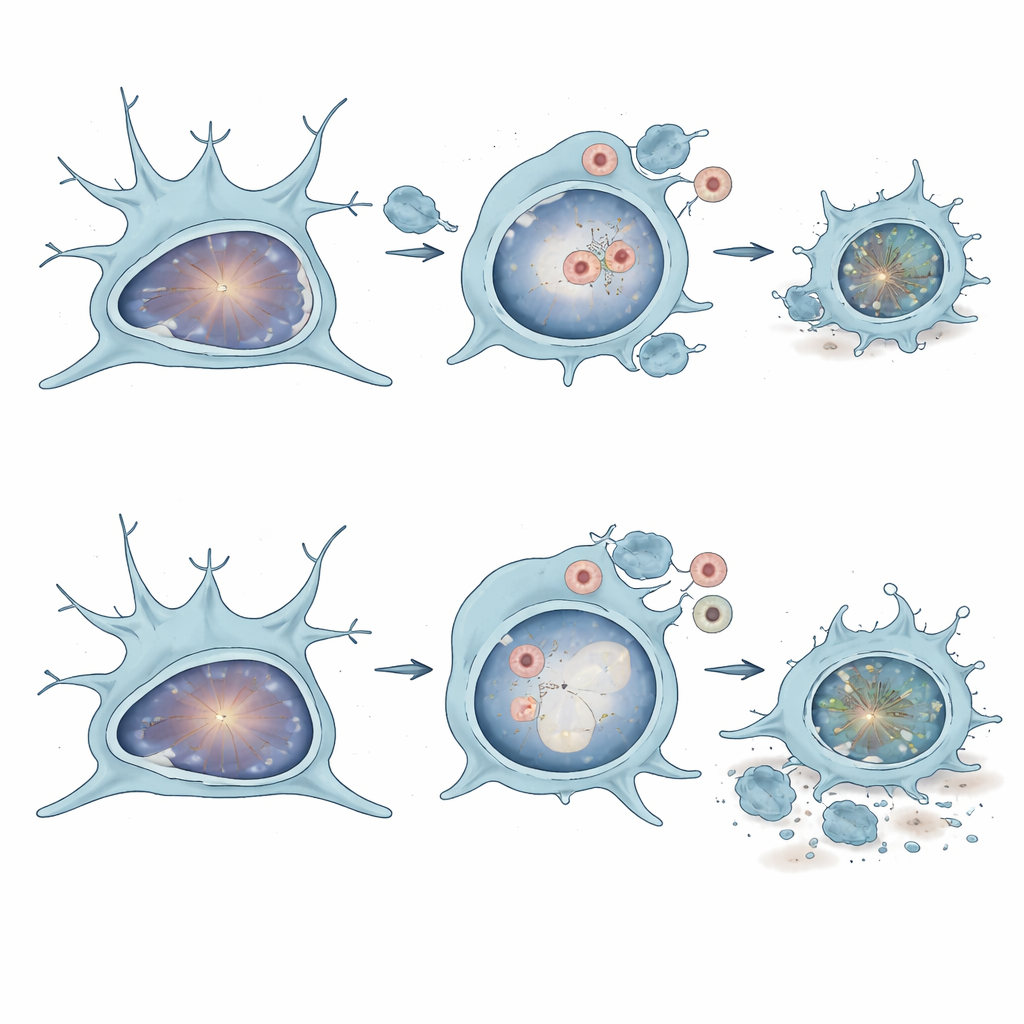
To test cathepsin B’s function, the researchers reduced or removed this enzyme specifically in myeloid lineage cells, which include microglia, in zebrafish, and knocked it out in all cells in mice. In both animals, microglia lacking normal cathepsin B activity accumulated large, poorly digested vacuoles filled with dead material. Live imaging in zebrafish revealed that these microglia formed more phagocytic compartments but fewer of them became properly acidified, suggesting that the “stomach” of the cell was not working efficiently. In mouse microglia grown in culture, those without cathepsin B initially acidified engulfed material but then rapidly lost their ability to maintain digestion over time, consistent with a progressive overload of undigested corpses.
Consequences for Circuits and Behavior
The failure to digest dying neurons had visible consequences in the developing brain. In zebrafish with impaired cathepsin B, the researchers saw more dead cells lingering both inside and outside microglia in the optic tectum, a key visual and motor center. These fish later showed abnormal locomotor behavior, swimming farther and faster than their normal counterparts. In mice lacking cathepsin B, the scientists found extra apoptotic cells specifically in the deep layer of the somatosensory cortex during a critical developmental window, along with more debris inside microglia. By adolescence, a particular class of excitatory neurons in these layers was reduced in number, and the animals displayed tactile hypersensitivity when their whiskers were gently stimulated.
What This Means for the Developing Brain
Taken together, the findings paint cathepsin B as a key part of the brain’s early-life housekeeping toolkit. Microglia without this enzyme can still find and engulf dying neurons, but they struggle to digest and clear them, leading to a buildup of debris, altered microglial shape and movement, and ultimately changes in how neural circuits are wired and how animals behave. While cathepsin B has sometimes been viewed as harmful in disease settings, this work suggests that, during development, it is essential for healthy brain maturation. Subtle disruptions in this kind of cellular cleanup machinery could therefore contribute to neurodevelopmental conditions where circuit formation and sensory processing go awry.
Citation: Silva, N.J., Anderson, S.R., Mula, S.A. et al. Microglial cathepsin B is necessary for neuronal efferocytosis in zebrafish and mice during brain development. Nat Commun 17, 3881 (2026). https://doi.org/10.1038/s41467-026-70350-1
Keywords: microglia, brain development, efferocytosis, cathepsin B, neuronal apoptosis