Clear Sky Science · en
Directional dynamics in the entorhinal cortex of male mice driven by behavioral constraints
How the Brain Knows Which Way It’s Pointing
Finding our way through the world depends on an inner sense of direction, built from brain cells that fire when an animal faces a particular way. This study asks a deceptively simple question: are those “compass” cells hard-wired, or can they change their role when the way we move through the world changes? By watching thousands of neurons in a key navigation area in mice, the authors reveal that much of this directional code is surprisingly flexible and shaped by experience.
A Brain Area That Maps Space
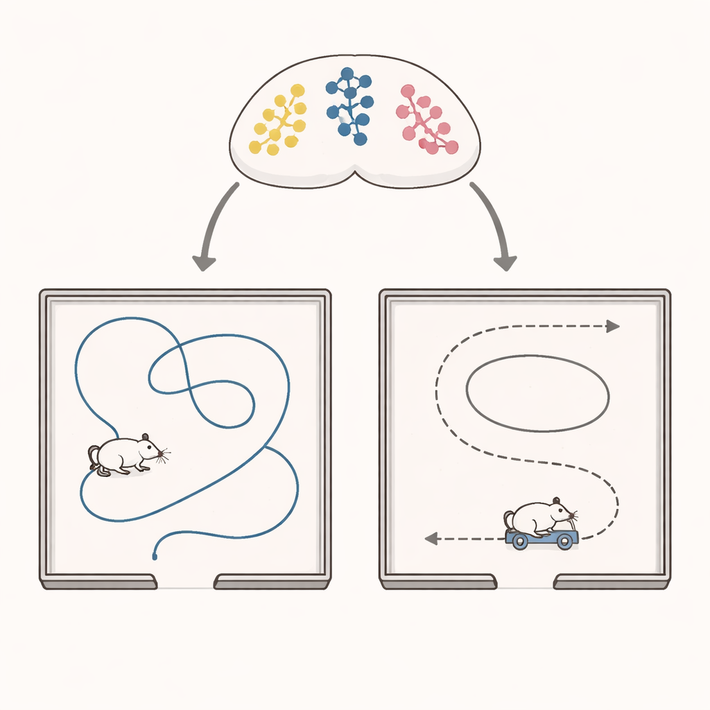
Deep in the brain, the medial entorhinal cortex works as part of a navigation hub. It hosts several kinds of specialized cells, including grid cells that map location and head-direction cells that care about which way the animal is facing. Until now, scientists did not know whether a cell’s “job description” as a head-direction cell was fixed or could be reassigned. The authors used two-photon calcium imaging to monitor over 11,000 neurons in mice as they explored a square arena. In some sessions the animals roamed freely; in others, their heads were fixed to a small autonomous cart that drove them around the same space, changing only how they moved, not where they went.
When Free Roaming Becomes a Guided Ride
The researchers first compared directional signals during free movement and cart-driven “assisted navigation” in a cue-rich arena with strong visual and odor landmarks. Surprisingly, overall direction tuning in the entorhinal cortex became sharper, more informative, and more stable when the mice were on the cart. But this improvement hid a striking reorganization. One group of neurons that clearly tracked head direction during free exploration lost this tuning on the cart. A second group, previously unremarkable, gained strong directional tuning only during assisted navigation. A smaller third group maintained reliable tuning in both conditions. Decoding analyses confirmed that the population signal for heading worked best when the decoder was trained and tested in the same navigation mode, showing that the pattern of active cells truly reconfigured.
Constraints, Cues, and a New Map
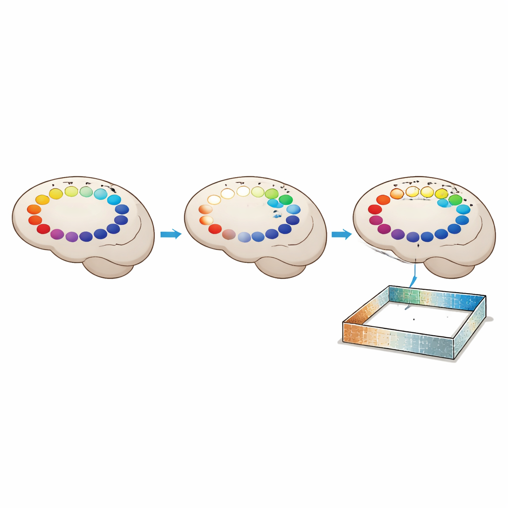
To probe what drives this switch, the team altered the environment and the cart’s movement. In a “cue-poor” arena stripped of most visual structure, assisted navigation no longer boosted directional coding: fewer cells gained tuning and maps were less stable. Changing the cart’s speed profile, however, made little difference; the new head-direction cells kept similar preferred directions even when the cart moved slower or faster. This points to rich external sensory cues, not simple motion statistics, as key ingredients for recruiting new directional cells under constraint. At the same time, the subclass of invariant cells preserved their coordinated firing across all conditions, suggesting they form a more hard-wired, attractor-like backbone for direction.
Learning a Second Compass
The authors then asked how quickly this assisted-navigation code forms. In mice experiencing the cart for the first time, the quality of directional maps, and the ability to decode heading from them, improved steadily over the course of a single session lasting only minutes. In later, “trained” sessions, these measures were already high and showed little change, implying that the new code had been learned and stored. During assisted navigation, many head-direction cells also began to carry information about where in the arena the animal was, not just which way it faced. Their spatial firing fields tended to cluster near walls, and their preferred directions pointed toward the nearest wall more often in the cue-rich than in the cue-poor environment. This indicates that, under behavioral constraints, the entorhinal map links direction and place through nearby sensory landmarks.
A Flexible Inner Compass
For a non-specialist, the central message is that the brain’s internal compass is not a single fixed mechanism. Instead, it mixes a small, stable core of direction cells with a larger, adaptable pool whose roles can switch depending on how the animal moves and what it senses. When head movements are constrained but rich cues are available, new neurons are recruited to help maintain a reliable sense of direction, and they quickly learn to tie that sense to specific locations and walls. This work suggests that our navigation system can store multiple maps for the same place, selecting the one that best fits the current way we move through the world.
Citation: Liu, R., Hao, J., Zhang, X. et al. Directional dynamics in the entorhinal cortex of male mice driven by behavioral constraints. Nat Commun 17, 3679 (2026). https://doi.org/10.1038/s41467-026-70289-3
Keywords: head direction cells, entorhinal cortex, spatial navigation, sensory cues, neural plasticity